Camptodactyly
1 Former Head of Hand Surgery Department, Catholic Children's Hospital Wilhelmstift, Hamburg, Germany
2 Board of PULPe (Pediatric Upper Limb Project Europe)
Abstract
Camptodactyly is a non-traumatic painless flexion contracture of a finger with the little finger most commonly affected. The exact cause is unknown.
Camptodactyly is classified into three types. The infantile type appears in infants and the juvenile type before puberty. The third syndromic type is part of distal arthrogryposis or certain syndromes and involves multiple fingers.
Several anatomical structures may be altered, particularly the flexor digitorum superficialis tendon, the retinaculum cutis and occasionally an atypical lumbrical muscle. These changes can cause shortening of the palmar skin, weakening of the central slip, and deformation of the bones around the PIP joint. When the contracture reaches 60 degrees or more, grasping is significantly impaired.
Early treatment is important to prevent secondary changes. Conservative management with well-fitted splints, manual therapy and good cooperation from both child and parents can often improve the contracture. If there is no improvement with conservative therapy despite good compliance, a stepwise palmar release operation can be indicated. During surgery, it is crucial to address the individual pathologies, as well as to follow up with thorough, intensive hand therapy after the operation.
Keywords
camptodactyly, flexion contracture, lumbrical muscle anomalies, palmar releaseIntroduction
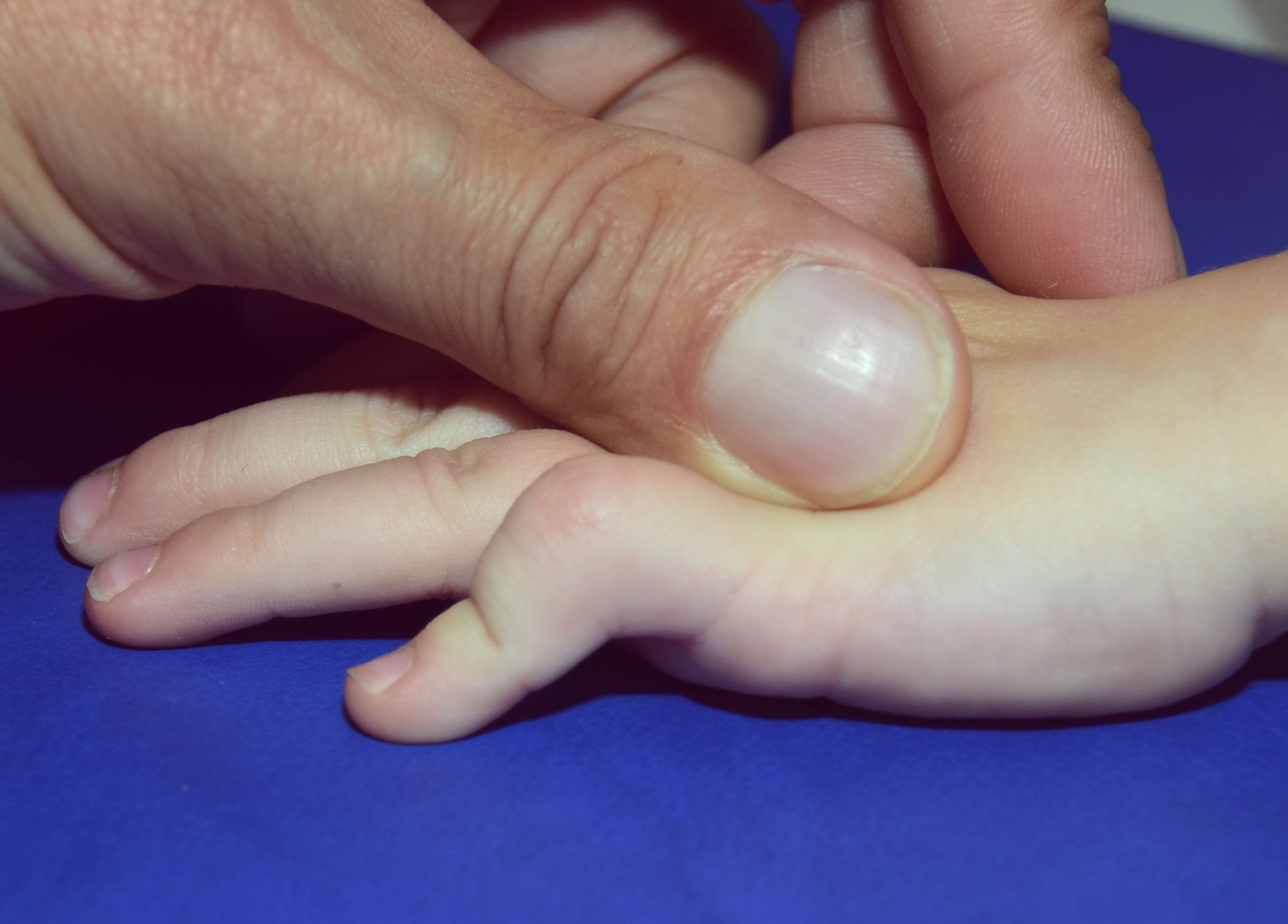
The term camptodactyly refers to a painless bending of the proximal interphalangeal joint (PIP), without traumatic aetiology (Figure 1). The term is derived from the Greek (kampto = I bend, daktylos = finger). There are two types: reducible forms, where only the active extension of the PIP is reduced, and fixed forms, where even passive extension cannot be achieved. Most cases are of the fixed form, with the contracture typically worsening gradually as the child grows.
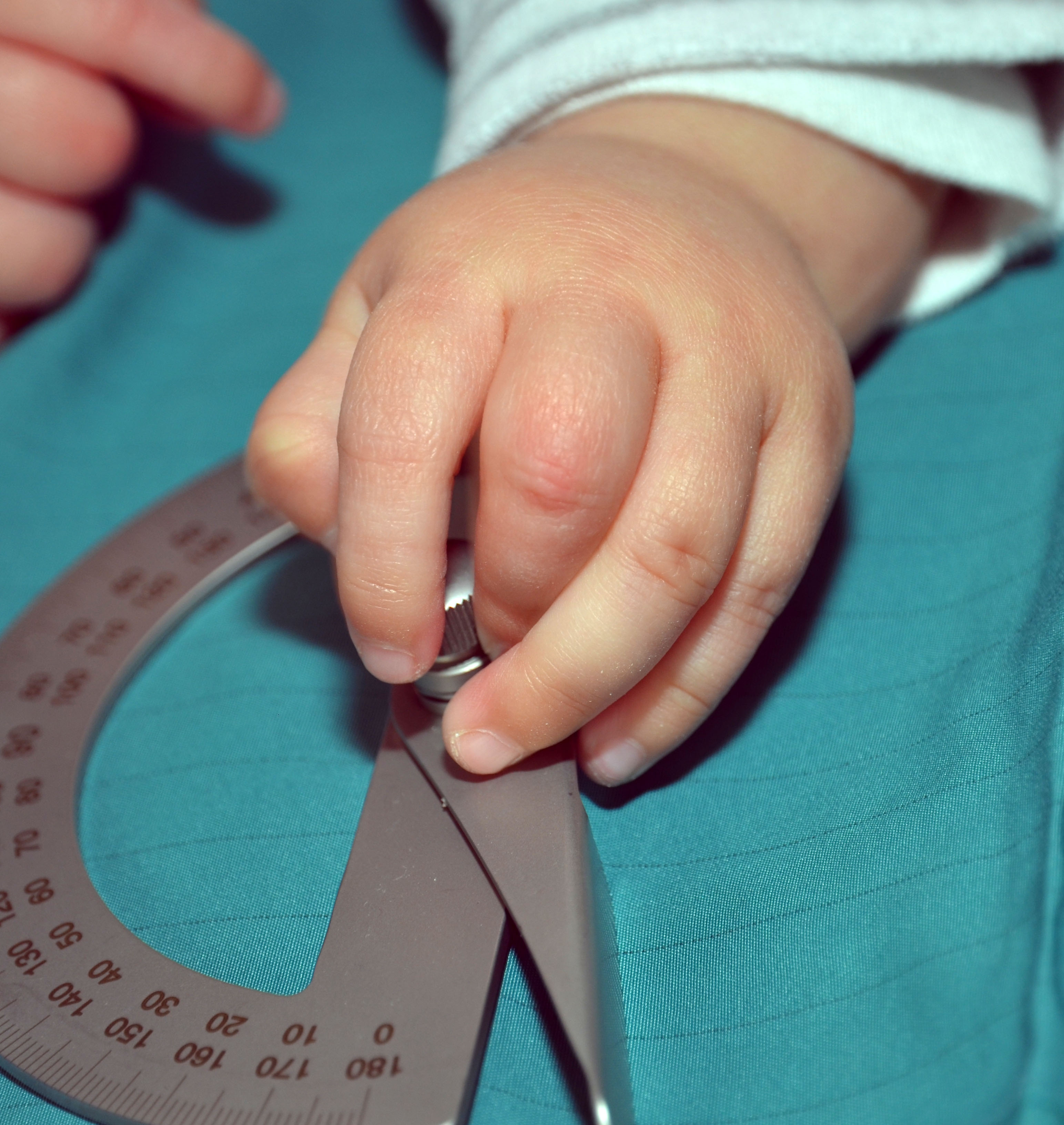
The resulting reduced range of motion (ROM) in the PIP joint affects hand function, making gripping difficult in more severe cases (Figure 2). While the metacarpophalangeal (MCP) and distal interphalangeal (DIP) joints are not directly involved, they may become misaligned as a means of compensation.
Basics
Epidemiology
Campodactyly affects less than 1% of the population and is bilateral in about two-thirds of cases. The little finger is most frequently involved [1], although other digits may also be affected. The incidence decreases towards the radial side of the hand.
Aetiology
The cause is unclear, but it is likely to result from an imbalance between the extensor and flexor muscles of the finger. While it usually occurs sporadically, it can also be inherited, typically following an autosomal dominant inheritance pattern with variable expression and incomplete penetrance [2].
Pathophysiology and anatomy
Various anatomical structures may be altered leading to flexion contracture. Primary abnormalities include the flexor digitorum superficialis tendon (FDS) and lumbrical anomalies, as well as a shortened retinaculum cutis. Secondary abnormalities involve palmar skin shortening, weakness of the central slip, palmar plate contracture of the PIP joint and dysplasia of the proximal phalangeal head [3]. It is believed that these secondary changes develop as a consequence of the extension deficit during growth.
An atypical insertion of the lumbrical muscles can contribute to flexion contracture when the muscle attaches to the FDS tendon proximal to the A1 pulley, thereby preventing the tendon from gliding beneath the pulley. The third and the fourth lumbrical muscles show a high incidence of anatomical variants. It is, however, debatable whether these variants contribute to contracture since they are more the rule than the exception [4], [5]. The lumbrical muscles normally originate on the radial side of the flexor digitorum profundus (FDP) tendon of the corresponding finger, run palmar to the deep metacarpal ligament and insert into the radial lateral band of the extensor apparatus at the level of the proximal phalanx. In doing so, they couple the FDP tendon to the extensor mechanism, the flexor forces are reduced in favour of increased extension. The 4th lumbrical muscle, which runs to the little finger, often originates bipennate at the 4th and 5th FDP tendon. In addition to the insertion at the lateral band, additional insertions have been found at the deep metacarpal ligament, the palmar plate of the MCP joint and at the bone of the proximal phalanx [5].
The interosseous muscles may also contribute to camptodactyly if they attach to the proximal phalanx [4], [6]. In the little finger, the third palmar interosseous muscle typically radiates into the radial lateral band of the extensor apparatus.
Classification
Types of Camptodactyly
Camptodactyly is classified into three types [7].
Type 1 infantile form: this occurs in infancy, is limited to the little finger and affects boys and girls equally. It is the most common form, affecting 84% of cases [8].
Type 2 juvenile form: this develops in prepubertal age (usually around 8 to 10 years of age) and mainly affects girls. It occurs in 13% of cases.
Type 3 syndromic form: frequently, several fingers in both extremities are affected and the contracture is often more pronounced and fixed than in forms 1 and 2. It is associated with a variety of syndromes of “arthrogryposis” disorders characterized by craniofacial deformities, short stature or chromosomal anomalies. Hand surgeons are more likely to see patients with milder cases such as distal arthrogryposis, Freeman-Sheldon syndrome, and Beals-Hecht syndrome [6].
Severity
Severity of camptodactyly is categorized by the degree of active PIP extension deficit as:
| Mild: | PIP extension deficit of <30 degrees |
| Moderate: | PIP extension deficit of 30–60 degrees |
| Severe: | PIP extension deficit of over 60 degrees |
Evalutation
The results are usually evaluated according to Siegert [9]:
| Excellent: | full extension with <15° loss of PIP joint flexion |
| Good: | correction to less than 20° of complete extension or more than 40° of increase in PIP extension with less than 30° of loss of flexion |
| Fair: | correction up to 40° of complete extension, or >20° increase in PIP extension with <45° flexion loss |
| Severe: | less than 20° improvement in PIP joint extension, or less than 40° of total PIP joint motion |
Diagnostics
Clinical examination
The position of the finger joints and their active and passive mobility are measured with the wrist held in a neutral position noting whether a firm or elastic resistance is felt during extension. The active extensor deficit (ED) and the passive ED (flexion contracture) are noted.
A careful examination of the hand often provides information about the underlying pathology [6], [7].
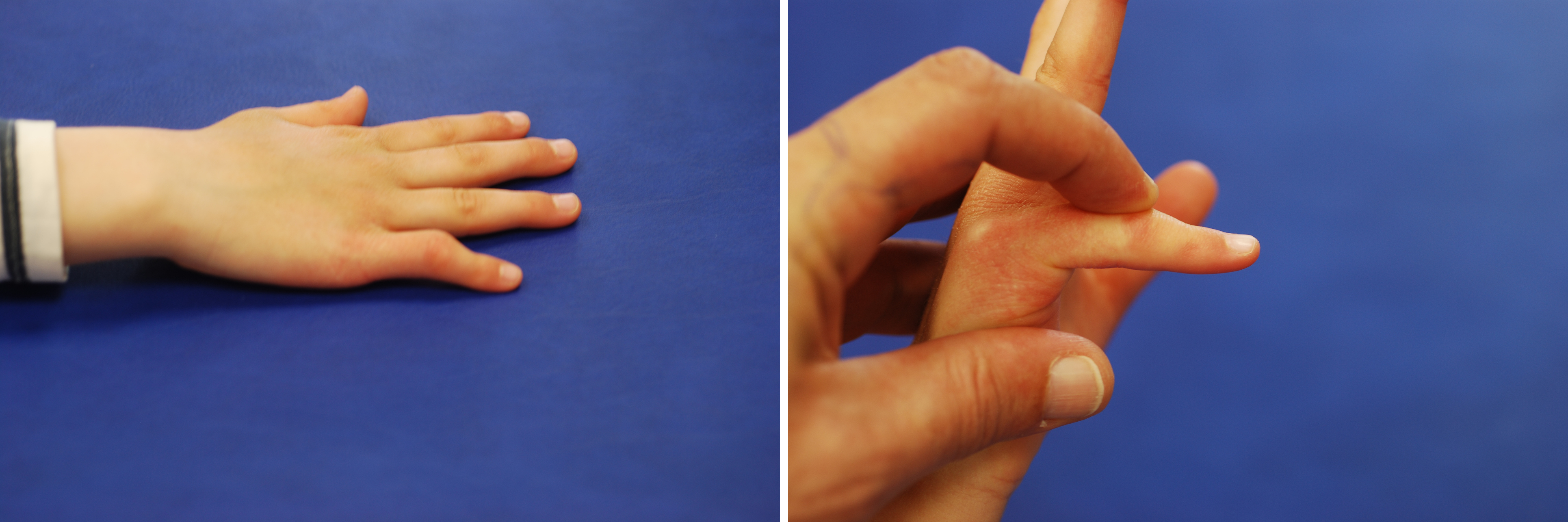
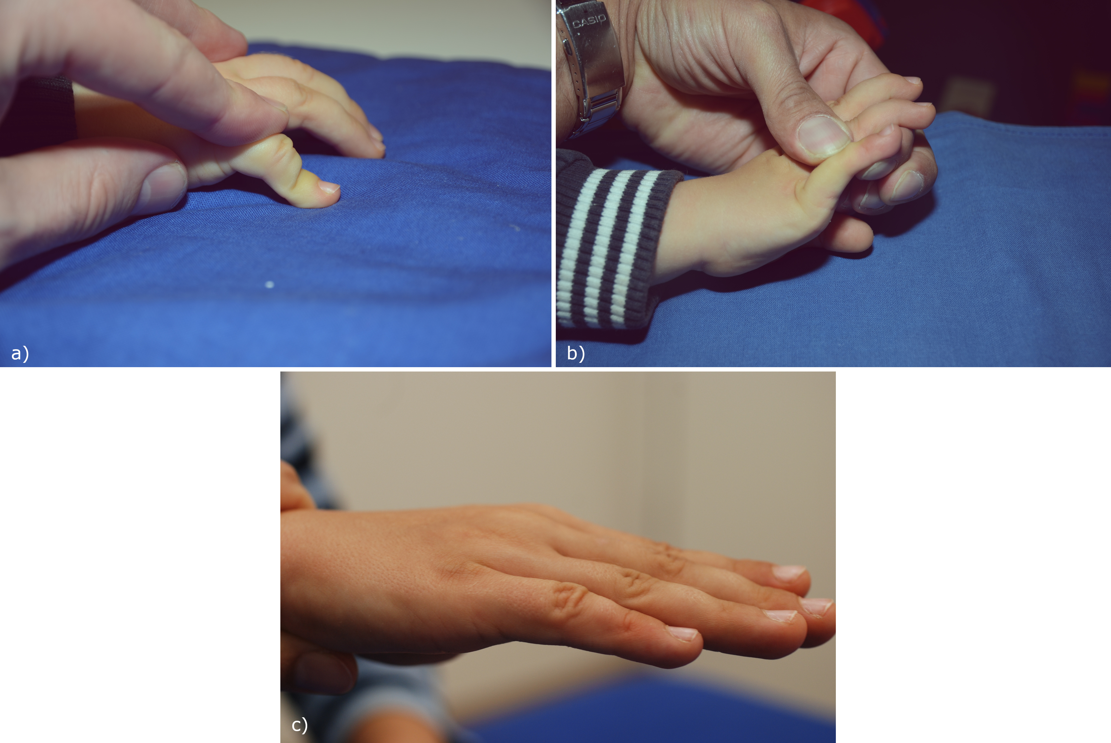
- Bouvier test (Figure 3): Compensatory hyperextension of the MCP is often found in camptodactyly. In the Bouvier test, the examiner places the MCP in slight flexion. If the PIP joint then can be actively extended, the test is positive. In camptodactyly, this may indicate lumbrical anomalies.
- Tenodesis test: The MCP joint is held alternately in extension and in flexion. If passive MCP flexion improves extension in the PIP joint, the causes of camptodactyly lie outside of the PIP joint.
If the Bouvier test is negative, there are three possible reasons:
a) Weakness of the elongated central slip, as in the case of long-standing flexion contracture. This is tested by the Extensor tenodesis test. Normally, the PIP is passively extended by traction of the extensor tendon when the wrist and MCP are fully flexed. If this does not happen, the central slip is attenuated.
b) Flexion tenodesis test indicates shortening of the FDP. In this case, extension becomes possible if MCP and wrist joint both are moved from the neutral position into flexion.
c) Arthrogenic contracture, i.e. shrinkage of the palmar plate of the PIP joint. If this is the case, extension of the PIP joint is not possible even when the MCP and wrist joint are held in flexion.
X-ray examination
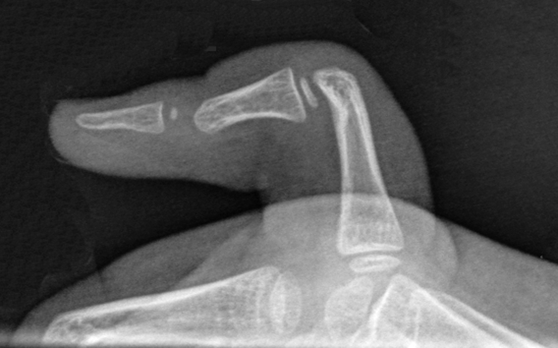
In moderate and severe camptodactyly, radiography of the affected finger is taken from the lateral view. Before the age of 3 years the ends of the phalanges consist mainly of cartilage and cannot yet be adequately assessed. Therefore, radiographs are not useful in young children. In severe contractures, the proximal phalanx loses its round, convex joint contour and it flattens and becomes beak-shaped due to palmar inclination of the neck (Figure 4). In severe contractures, the middle phalanx is subluxated palmarly and can create an indentation in the palmar neck of the proximal phalanx. In children with passively extendable PIP joints, the condyles are better formed than in fixed contractures.
Therapy
The preoperative findings are crucial for the treatment. Important factors are the age of the child, the extent of the PIP contracture and whether the contracture can be reduced or not.
Conservative therapy
Treatment for all affected individuals begins with conservative therapy by manual stretching and serial splinting. Splints are used for flexion contractures of around 40° or more. Especially in toddlers and preschool kids (Benson's type 1 camptodactyly), the contractures often respond very well. Furthermore, the compliance of patients and their relatives is checked, as surgery can only be successful if postoperative hand therapy is carried out reliably. Korean authors have achieved an improvement in the extension deficit in children under 3 years of age by means of manual stretching alone, regardless of the degree of severity: from 20° to 0° in mild forms, from 40° to 10° in moderate forms, and from 75° to 30° in severe cases [10]. The effectiveness of splints has also been demonstrated by Japanese authors. They treated children aged 10 months to 8 years with a dynamic finger extension splint with lateral spiral springs (Capener splint), which was worn for 24 hours a day for several months. This achieved full to greatly improved extension. However, the contractures tended to deteriorate again at the end of therapy [11].
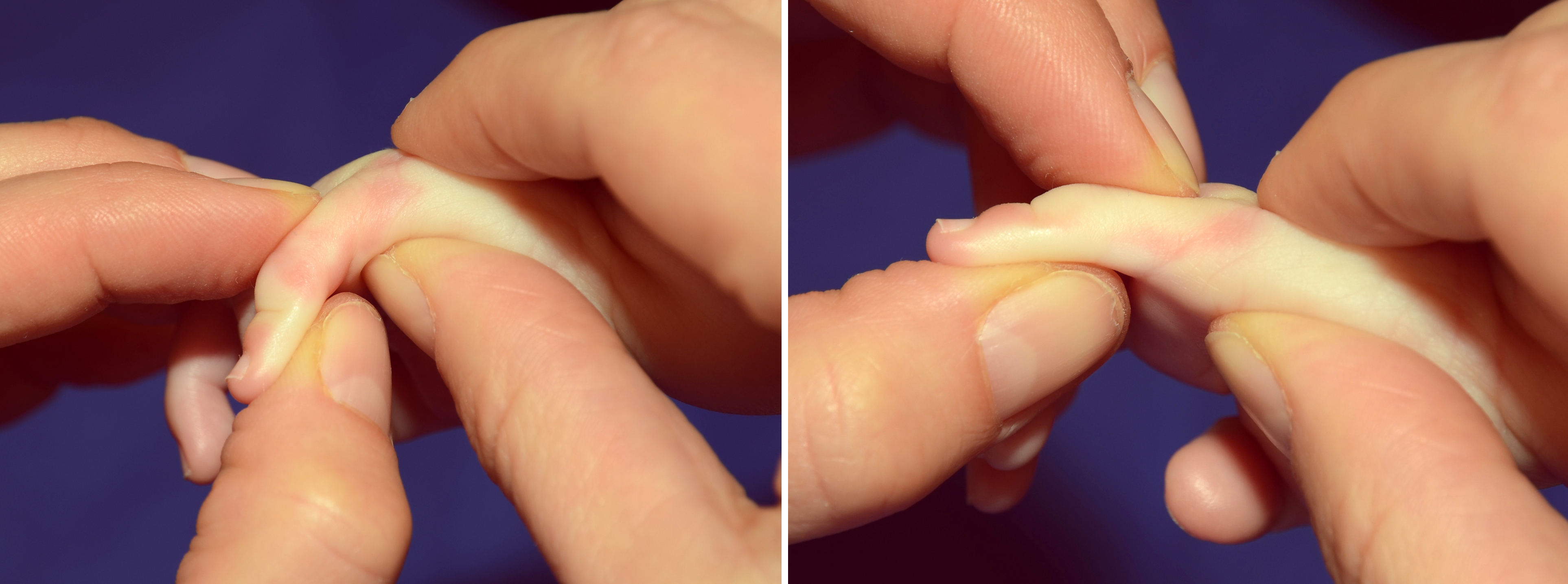
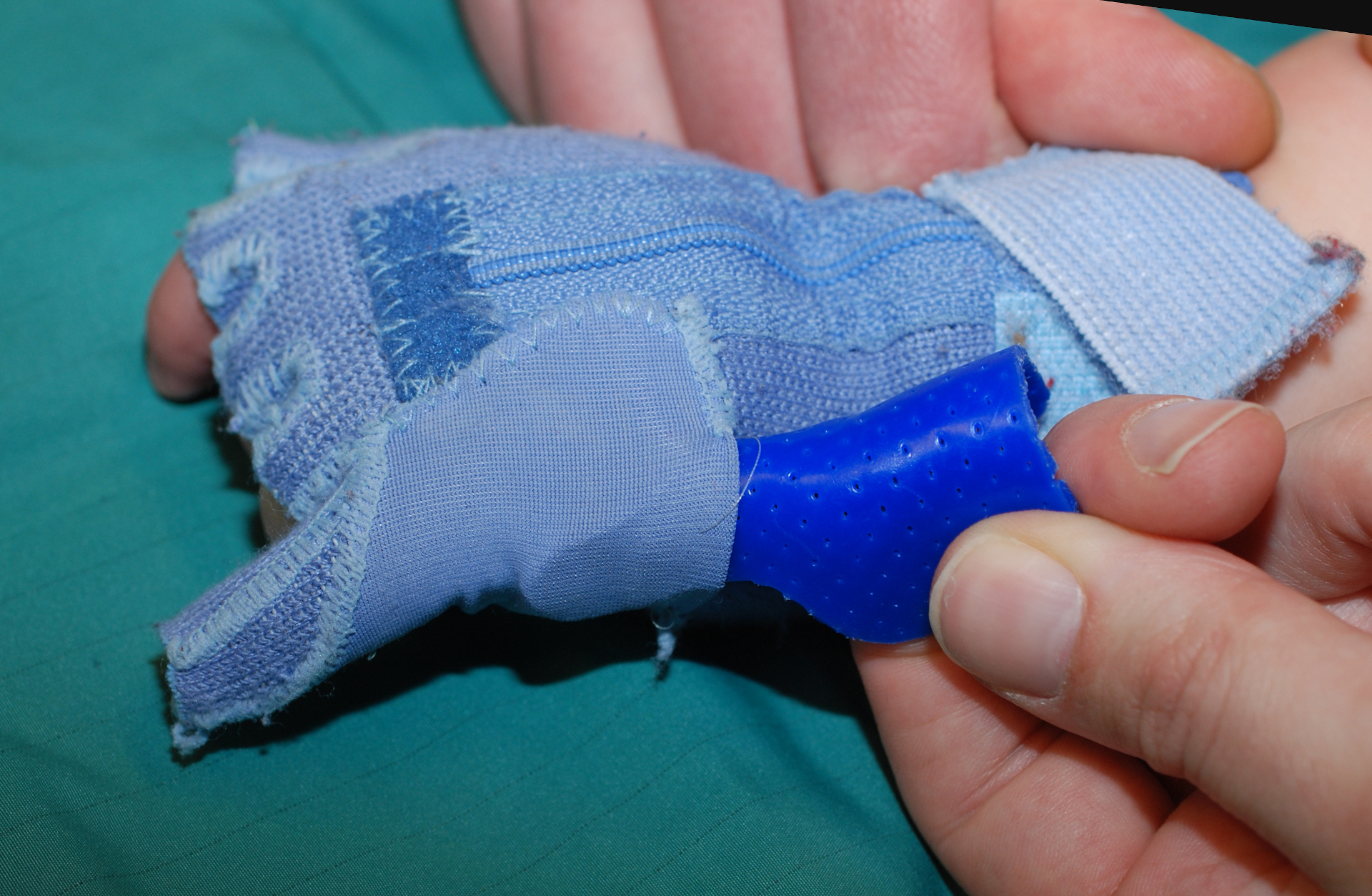
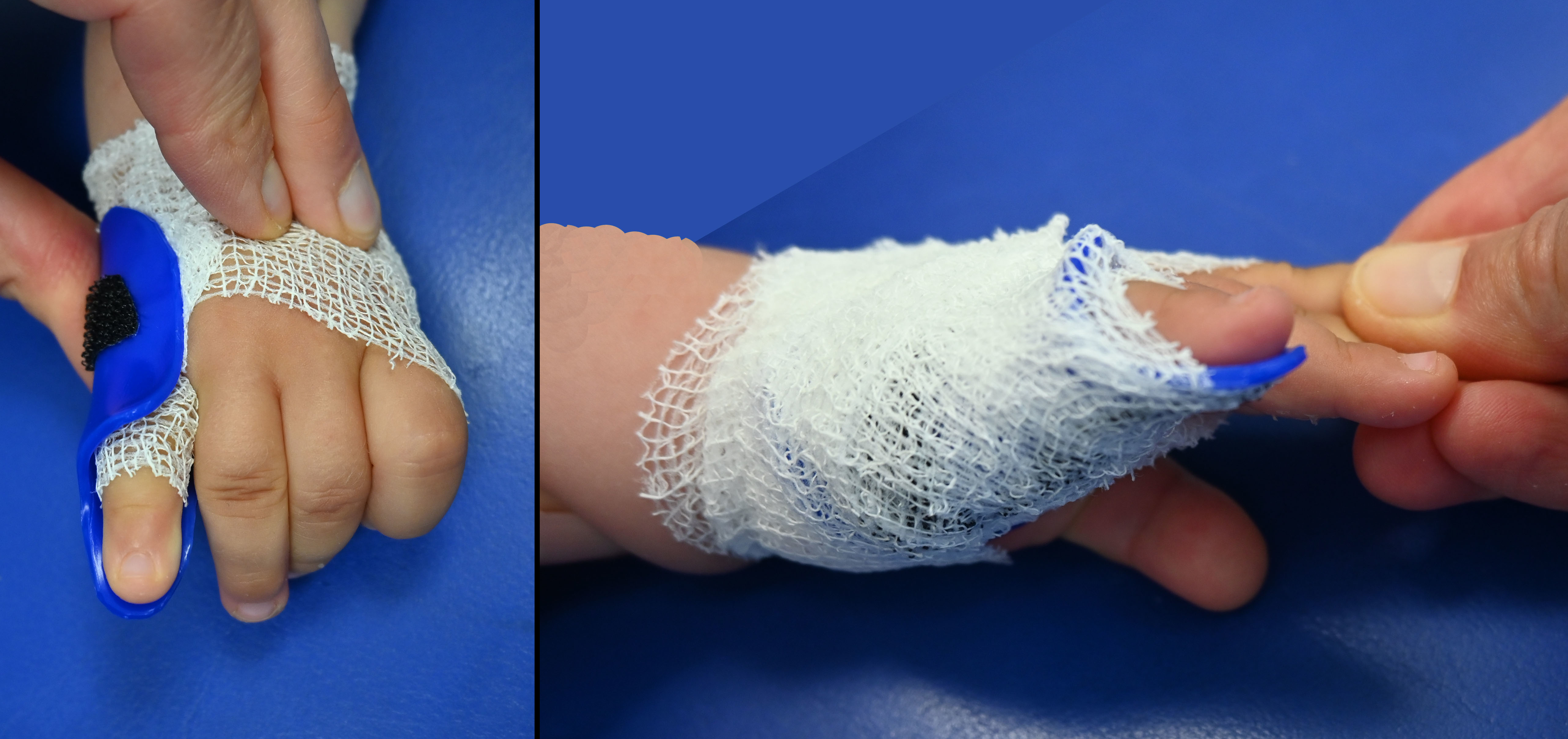
Splint treatment should begin as early as possible and can be implemented from the age of 8 months. The challenge lies in creating well-fitting splints for children who are unable to remain still. For children under 3 years of age, static splints such as the Glove Splint can be used to stretch the finger (Figure 7). Some parents found it difficult to put on the tight glove. We have therefore developed a simpler solution to prevent the thermoplastic splint from slipping by using a cohesive elastic fixation bandage instead of the glove (Figure 8).
For older children, dynamic extension splints may be used several times an hour during the day combined with static splints worn at night (Figure 9).
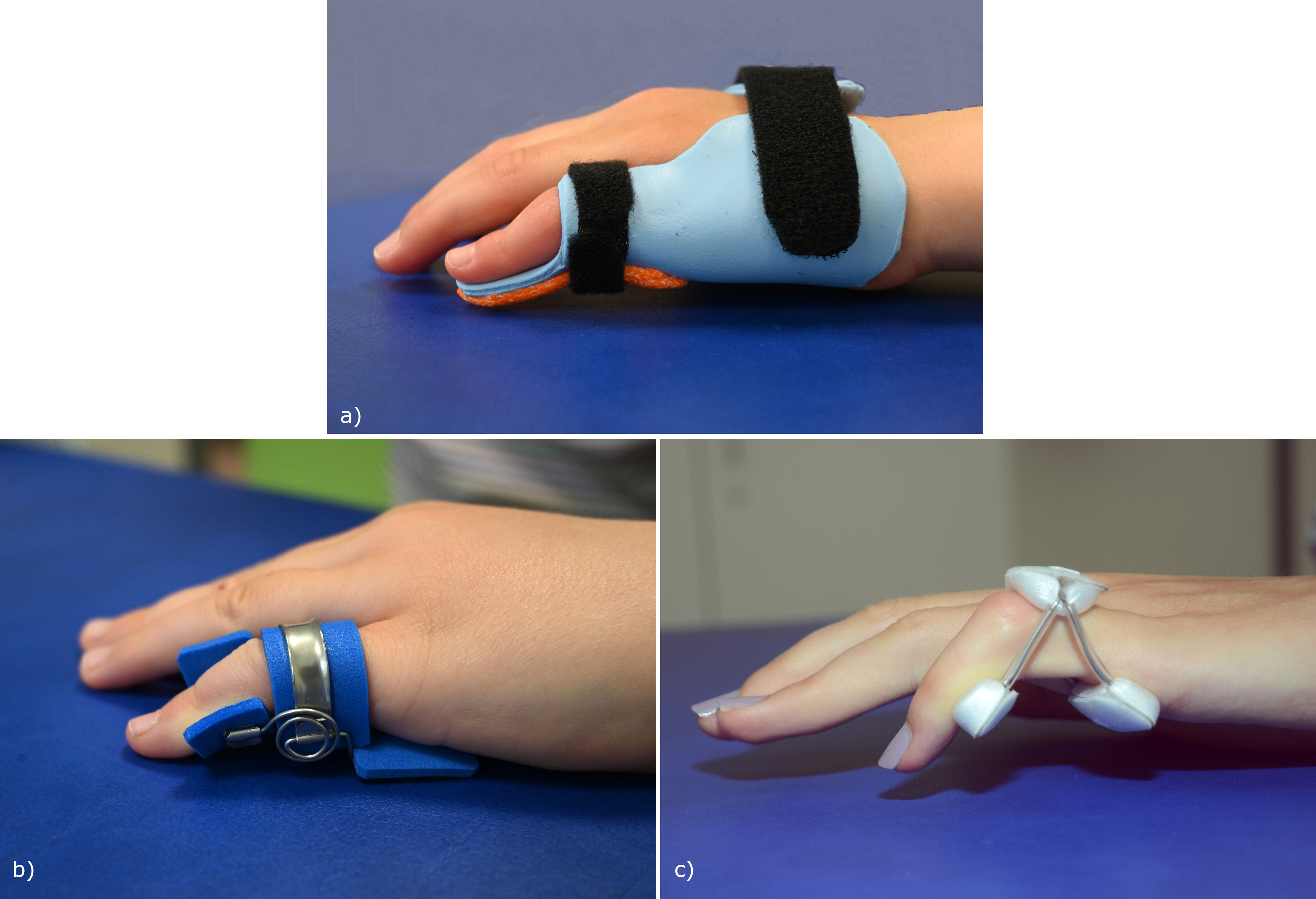
In adolescents, in addition to manual stretching, prefabricated dynamic finger splints are used.
We recommend wearing the dynamic finger extension splints during the day for about 30–60 minutes, 3–5 times a day and supplementing it with thermoplastic night splints.
It is essential for children to attend regular follow-up appointments to monitor compliance and assess both the fit of the splints and the condition of the skin. Adjustments to the splints should be made as needed. The deformity typically worsens during growth spurts, especially during the periods of rapid growth between 1 and 4 years of age and 10 and 14 years of age. A deterioration is a reason to start splint therapy again and intensify stretching.
Operative Therapy
Splinting is mandatory before planned surgical interventions for camptodactyly in order to test compliance. A lack of cooperation is a contraindication for surgery [12], since the success depends largely on the postoperative hand therapy, which can be uncomfortable and requires the patient's motivation and cooperation for several months.
Surgery is indicated
- if hand function is impaired. The compensatory MCP hyperextension hinders function, affects writing, and can interfere during ball sports.
- after unsuccessful, consistent use of an orthosis for at least 6 months
- if the extension deficit remains above 60°. (Some surgeons already operate on milder cases with an extension deficit of 30 to 55°)
- in case of bony changes, but only if the PIP joint is not completely dislocated palmarly or the joint is not destroyed.
- in pronounced and progressive camptodactyly in the young child
Simple camptodactyly
If the PIP joint is primarily flexible or if the contracture has improved with splint therapy and has become flexible, the Bouvier test is performed. If the Bouvier test is positive, i.e. the PIP joint can be actively extended when the MCP is held in slight flexion, there may be an anomalous insertion of the lumbrical muscle. Even if it is unclear which lumbrical insertion changes are pathological, they should be carefully examined through a sufficiently large incision and addressed by detachment of the anomalous insertion from the bone, from the MCP capsule, from the tendon sheath and pulley or from the FDS tendon. Such surgical revisions generally have a favourable prognosis.
Technique of stepwise release [13]:
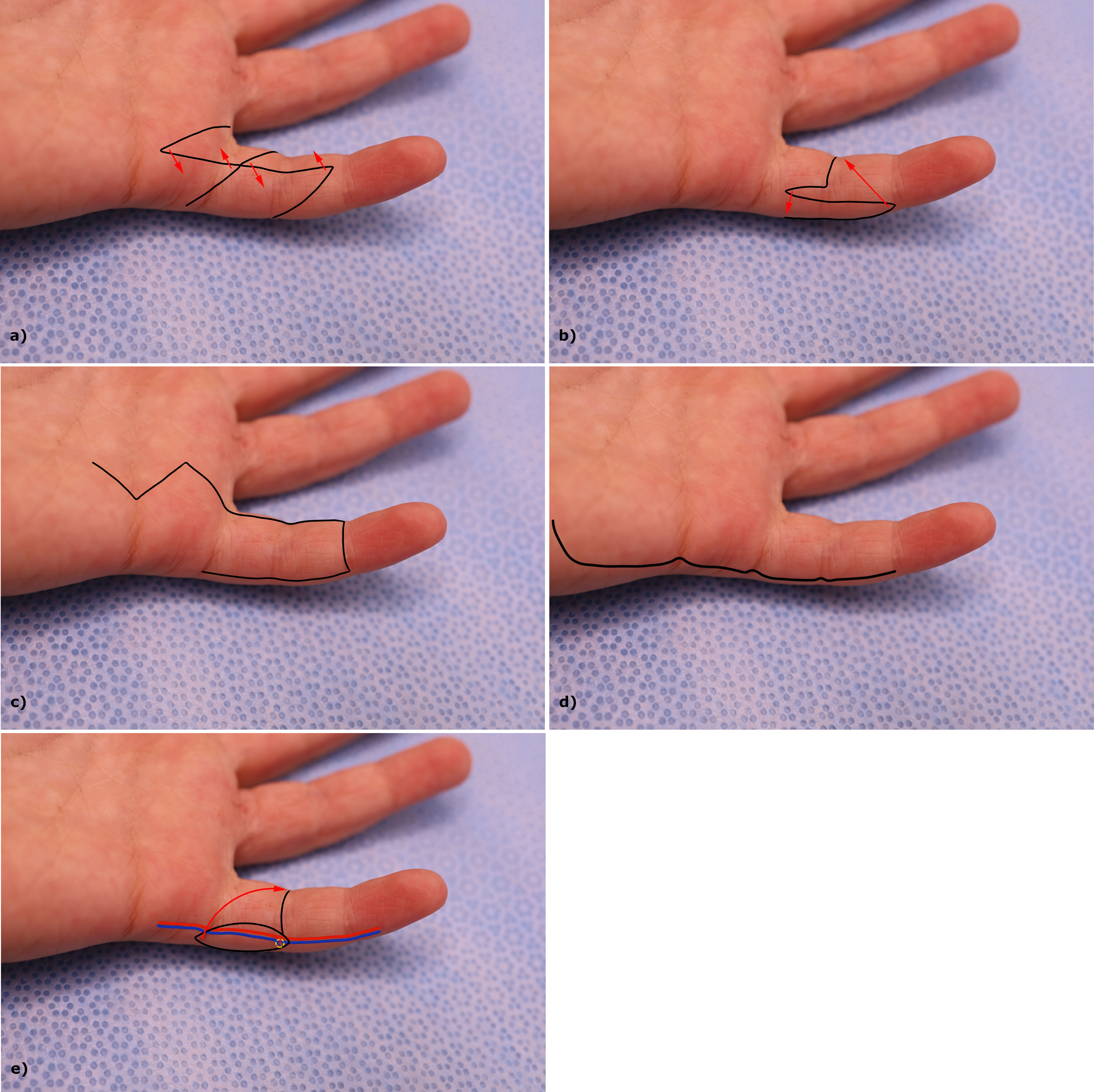
- Skin elongation: using a central longitudinal incision and multiple Z-plasties or a lateral transposition flap/ stiletto flap [14] or an extended Malek incision [15] or longitudinal lateral incision or digital artery perforator flap [16] (Figure 10)
- Release of subcutaneous tissue
- Check the FDS tendon for altered fibrous texture
- Check for an atypical lumbrical insertion and detachment in the case of an atypical one, e.g. on the bone, MCP capsule, sheath or FDS tendon
To place the MCP joint in slight flexion, some authors recommend an additional lasso procedure or transposition to the lateral band if the FDS tendon is functional. In the lasso procedure, the FDS tendon is wrapped around the A1 pulley after being detached [6], [12]. Others transpose the FDS tendon to the lateral bands of the extensor apparatus, in order to strengthen extension [17]. This step carries the risk of worsening flexion. Many malformation surgeons advise against it [18], [19].
Severe camptodactyly
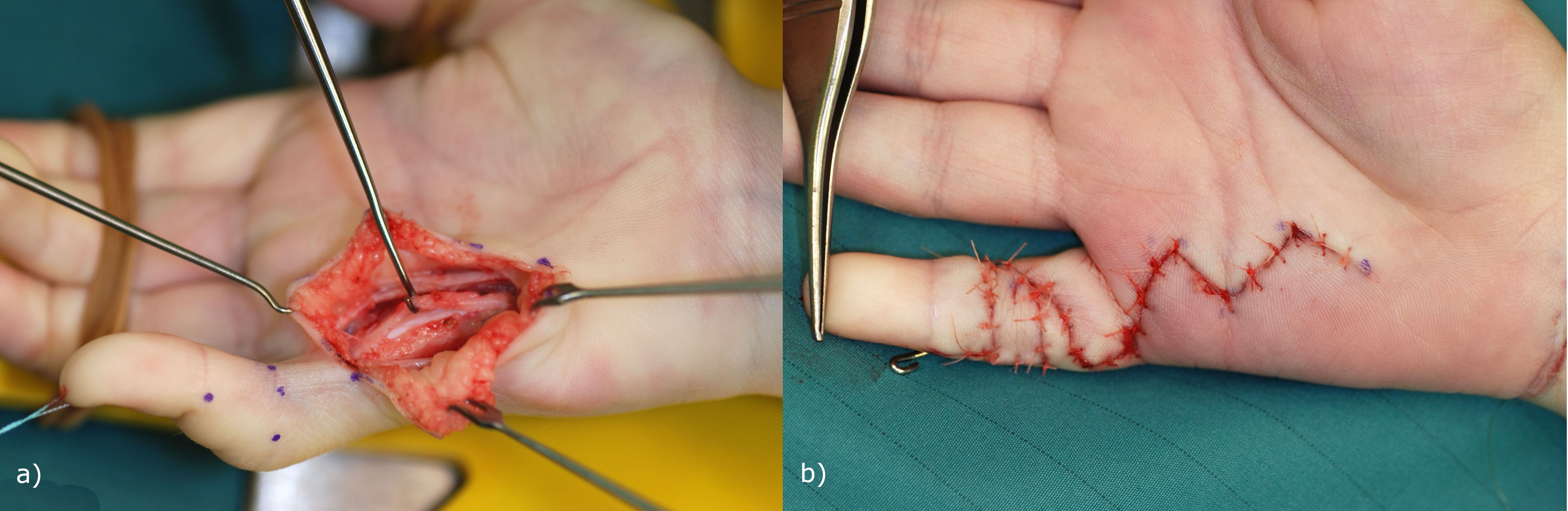
In severe forms further steps are taken to achieve a complete palmar release [13].
Technique for step-by-step palmar release
Step 1 to 4 as described above in simple camptodactyly.
- If the contracture persists: Open the tendon sheath at the distal edge of the A2 pulley, detach the altered FDS tendon at PIP level. The FDS tendon is usually shortened, rigid and inelastic. The transposition of an FDS tendon that has been modified in this way is rather functionless. Its transposition to the extensor side is not useful.
- If PIP contracture persists, perform a stepwise arthrolysis, beginning with transection of the check-rein ligaments.
- Check the extensor mechanism for weakness. Decide whether to use a post-operative MCP joint extension block splint or a relative motion flexion splint. The relative motion splint prevents hyperextension of the MCP joint during active extension exercises and allows targeted PIP extension exercises.
- Temporary PIP arthrodesis with K-wire for 3 weeks in a stretched position.
- Check blood supply in the finger. If the blood supply is compromised, correct the position of the PIP into a slightly more flexed position.
Postoperative management
Postoperative care, hand therapy and patient cooperation are crucial for a good surgical outcome.
After palmar release:
- The K-wire is protected with a splint, the PIP joint is relieved by a flexion position in the MCP joint, the wrist in neutral position. The distal phalanx remains free so that the FDP tendon can be moved immediately. After wound healing and soft tissue consolidation, the K-wire is removed 3 weeks postoperatively and hand therapy is started.
- Practice bending. A night splint in PIP extension must be worn for another 4 to 6 weeks and if needed, a finger extension splint (e.g. Ruck® splint or Capener splint) during the day.
- After 6 weeks, no daytime splint and only a night splint for around 4 to 6 months.
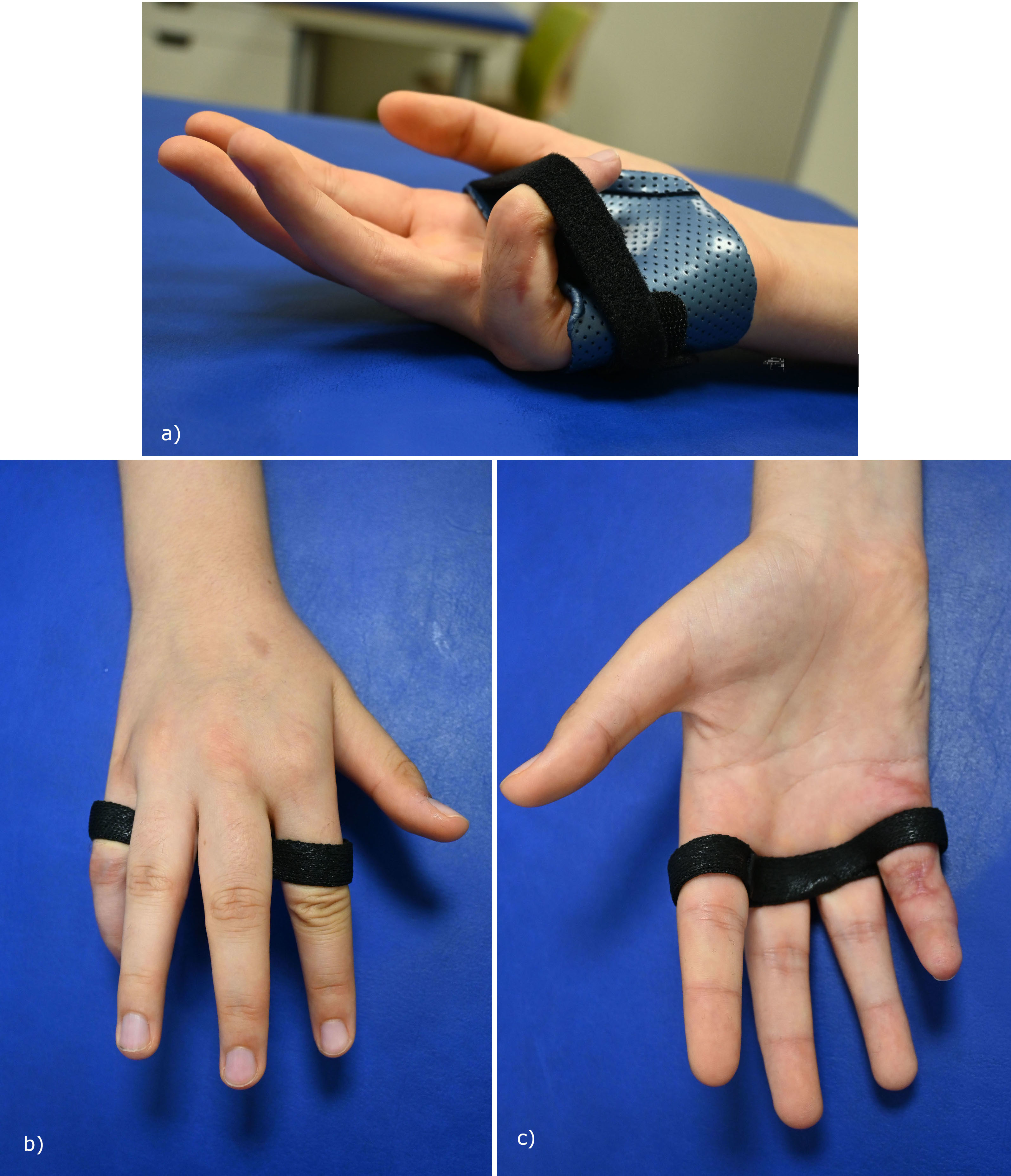
- If there are signs of weakness of the central slip an extension block splint or relative motion flexion splint (Figure 12) must be used. If active extension deficit is <60° in the extensor tenodesis test use it for 4 weeks and if the deficit is >60° some additional weeks.
The good results obtained using a treatment algorithm and a step-by-step intraoperative approach are encouraging [12], [20].
Complications
Early complications such as pin tract infection or graft healing problems are uncommon and should be addressed early. Digital nerves may be affected by too aggressive extension, potentially resulting in sensory disturbance. Stiffness is the most frequent complication and can affect both extension and flexion of the finger. A meta-analysis of the 16 most significant studies reported a low overall complication rate (2%–11%) [21], although this may be higher if the recommended protocols for surgery and rehabilitation are not followed.
Results
Camptodactyly is usually not a very disabling anomaly, and surgical intervention should not be undertaken lightly. The goal of the operation is to achieve a more extended position of the finger to enable grasping and bimanual tasks [14]. Normalization is not possible. Patients and parents must be thoroughly informed about the anticipated outcomes, the risk of finger stiffness and the importance of adhering to pre- and postoperative rehabilitation instructions and splinting. The surgical release is not a simple operation and requires careful preparation and experience. The risk of failed treatment with worsening of the camptodactyly is likely to be higher in less specialised centres than in those with more frequent camptodactyly operations and access to high quality hand therapy.
Treatment failure in older publications and after late correction of children with stiff PIP joints has been reported in 14% up to 65% [8], [12]. Most authors have found that the extent of active PIP joint mobility can be shifted into the extension range by surgery, but that total joint motion cannot be improved [22], [23]. This had led to the dogma that surgery should be avoided if possible. However, with sophisticated conservative or combined conservative and operative treatment the extent of flexion contracture in most patients can be reduced (Corain et al. mean from 65° extension lag to 16,5°; Netscher et al. from 62° to 4° [15], [19]). A shift in the PIP range of motion towards extension nowadays tends to mean both an improvement in finger function and in appearance. However, a drawback is that loss of strength due to reduced flexion of the little finger can impair power grip in the hand.
British surgeons have suggested early tenotomy of the FDS to reduce extension deficit in severe contractures in young children [20]. This could make conservative splint therapy possible and could perhaps prevent or reduce secondary attenuation of the central slip with worsening of the contracture with growth. In a follow-up of 2.5 years on average, the extension deficit was reduced from 30–90° to 0–60° [20].
It has been shown that bony deformities of the proximal phalangeal head can be reduced and even normalised after soft tissue surgery alone [15], [19].
Special challenges
FDS tenotomy to enable splinting
With the knowledge that the central slip becomes weaker with time in the case of a 60° contracture, early release is recommended by British surgeons [20]:
- if the 60° contracture persists after splinting and
- if the contracture rapidly progresses from 30° within a year.
The authors distinguish between two groups, those with “simple” and those with “complex” camptodactyly. In the case of “simple” camptodactyly (age under 10 years, one finger affected, not syndromal), the incision is made in the distal palmar crease and the FDS is tenotomised as distally as possible with the finger flexed, combined with careful passive release of connective tissue adhesions. In “complex” cases (patients aged 10 years or older, multiple fingers affected, syndromic patients, previous surgery), the Bruner incision is extended distally and the FDS tenotomy is performed at chiasma level with lumbrical examination. This is followed by careful splint treatment. The improved extension deficit makes conservative splint therapy possible even in the case of severe contractures in young children and can thus prevent progressive contracture with growth [20].
Type 3 syndromic camptodactyly
Camptodactyly is sometimes part of arthrogryposis disorders. In distal arthrogryposis, there is often a windblown deformity with ulnar deviation of the fingers in the MCP joints. Several fingers of both hands are affected (see chapter Distal arthrogryposis).
The contractures may be tenogenic or arthrogenic in origin. This can be easily verified clinically with the tenodesis test. If passive PIP extension improves when the wrist is placed in maximal flexion, the tenodesis test is positive, indicating a shortening of the extrinsic flexors.
Also, these contractures can be significantly improved by conservative therapy. However, the different flexion positions of the fingers require more elaborate splints in order to effectively stretch the individually flexed fingers (Figure 13, also see chapter Splints).
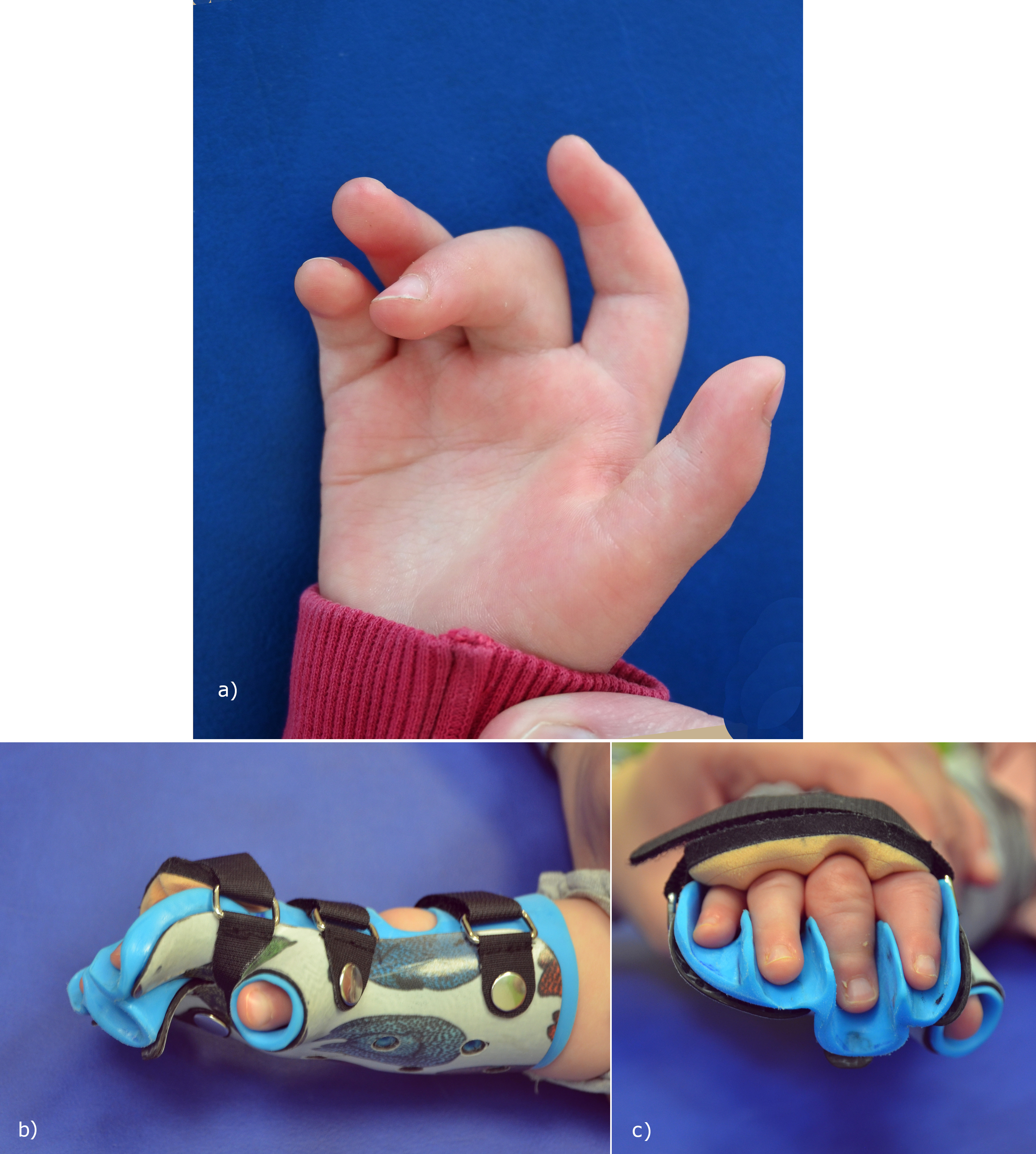
If in case of a tenogenic contracture splint treatment is not successful, and the extrinsic flexors may be lengthened surgically. This is possible at the forearm level by a Z-plasty of the tendon and suturing the cut ends in an elongated state or as a fractional lengthening at the muscle-tendon junction [24]. However, the lengthening weakens the grip. If not tenogenic, surgery may follow the recommendations for types 1 and 2 camptodactyly.
Salvage operation
If, in older children and adolescents, the fixed flexion contracture with only minimal residual mobility significantly impairs function and there are severe bony joint changes or poor compliance, then a shortening closing-wedge osteotomy or PIP joint arthrodesis is a simple and safe solution as a salvage operation [25]. This approach is also applicable after previous procedures that have failed to sufficiently reduce the flexion contracture. Performing a PIP fusion in a more extended position can, in some cases improve hand function. Currently, the little finger PIP joint is set in the “keyboard position” of about 20° flexion, no longer in the “tool position” of 45°–50°, as it used to be. The patient's individual needs are important for decision making.
Author’s preferred method
We always start therapy with consistent manual stretching and splint treatment.
- For children under 3 years of age, static splints can be used for stretching, while for older children, dynamic extension splints can be used during the day and static ones at night.
- For toddlers with fixed contracture and lack of improvement with the splint treatment: we perform surgical severing of the FDS tendon to create a better position that allows splint treatment.
- In adolescents, in addition to manual stretching, dynamic finger splints are used (Figure 9 b, c). We recommend wearing the dynamic finger extension splints during the day for about 30–60 minutes, 3–5 times a day and supplementing it with thermoplastic night splints.
Operative therapy: There is no benefit to be gained from surgical treatment of flexion contractures of less than 30°.
Surgical treatment is indicated for flexion contractures of 60° or more, or 40° in some cases.
Another indication for a palmar release is bony joint changes.
Severe cases should be treated as early as possible to restore the disturbed balance and minimize secondary pathologies. Indicated in >60° contracture, if patients and families want it and cooperation is proven. The step-by-step palmar release respects the individual pathology. This customized approach addresses the structures responsible for camptodactyly specifically.
As a salvage operation, PIP arthrodesis in a more extended position can be an option for adolescents at the end of the growth period and for those who are lacking in compliance to treatment protocols.
Conclusion
Normalization of finger joint motion affected by camptodactyly is not possible. The goal of therapy is to improve hand function by reducing the contracture and preventing further deterioration. It is important to give the patient and their parents a realistic impression of the expected results before starting treatment, and to emphasize the importance of compliance to treatment protocols.
Conservative therapy with serial splints and manual stretching improves the extent of the flexion contracture in many patients and is the first therapy for all. In addition to improvement, it also serves to check cooperation. An absolute requirement for a surgical release is compliance in view of the lengthy and uncomfortable postoperative hand therapy. In the past, it was generally recommended to avoid surgery, to treat non-surgically with splints and hand therapy, and to operate only if there was a fixed PIP contracture of more than 60°. Nowadays, it's seen as important to distinguish the primary causes from the secondary consequences of delayed treatment. Identifying the individual pathologies by clinical examination and in the step-by-step intraoperative procedure is key to improving the results of surgery.
Severe cases should be treated as early as possible to restore the disturbed balance and minimize secondary pathologies. The surgical treatment must address all pathologies step by step. Transposition of a rigid, fibrosed FDS tendon to the extensor side is not useful. The transposition of the transected FDS to the extensor side also carries the risk of over-reinforcing the extensor apparatus.
The postoperative treatment is just as important for a good result as a careful operation.
All treated patients must be regularly monitored until the end of growth in order to detect deterioration and quickly re-start manual and splint treatment.
References
[1] Senrui H. Congenital contractures. In: Buck-Gramcko D. eds. Congenital malformations of the hand and forearm. London: Churchill Livingstone; 1998. 295-309[2] Welch JP, Temtamy SA. Hereditary contractures of the fingers (camptodactyly). J Med Genet.1966; 3:104–113. DOI: 10.1136/jmg.3.2.104
[3] Smith PJ, Grobbelaar AO. Camptodactyly: a unifying theory and approach to surgical treatment. J Hand Surg Am. 1998 Jan;23(1):14-9. DOI: 10.1016/S0363-5023(98)80082-8.
[4] Schmidt HM, Lanz U. Surgical Anatomy of the Hand. Stuttgart, New York: Thieme; 2004
[5] Eladoumikdachi F, Valkov PL, Thomas J et al. Anatomy of the intrinsic hand muscles revisited: part II. Lumbricals. Plast Reconstr Surg. 2002 Oct;110(5):1225-31. DOI: 10.1097/00006534-200210000-00002
[6] Lanz U, Foucher G, Habenicht R, Kall S, Schmidt H-M. Camptodactyly. In Hovius S, Foucher G, Raimondi L; Federation of European Societies for Surgery of the Hand, editors. The Pediatric Upper Limb. London: Martin Dunitz; 2002. p. 103-131
[7] Benson LS, Waters PM, Kamil NI, Simmons BP, Upton J 3rd. Camptodactyly: classification and results of nonoperative treatment. J Pediatr Orthop. 1994 Nov-Dec;14(6):814-9. DOI: 10.1097/01241398-199414060-00024
[8] Engber, WD, Flatt AE. Campotodactyly an analysis of sixty-six patients and twenty-four operations. J Hand Surg Am. 1977 May;2(3):216-24. DOI: 10.1016/s0363-5023(77)80072-5
[9] Siegert JJ, Cooney WP, Dobyns JH. Management of simple camptodactyly. J Hand Surg Br. 1990 May;15(2):181-9. DOI: 10.1016/0266-7681_90_90122-k
[10] Rhee SH, Oh WS, Lee HJ, Roh YH, Lee JO, Baek GH. Effect of passive stretching on simple camptodactyly in children younger than three years of age. J Hand Surg Am. 2010 Nov;35(11):1768-73. DOI: 10.1016/j.jhsa.2010.07.032
[11] Hori M, Nakamura R. Inoue G, Imamura T, Horii E, Tanaka Y, Miura T. Nonoperative treatment of camptodactyly. J Hand Surg Am. 1987, 12(6):1061-5. DOI: 10.1016/s0363-5023(87)80112-0
[12] Foucher G, Loréa P, Khouri RK, Medina J, Pivato G. Camptodactyly as a spectrum of congenital deficiencies: a treatment algorithm based on clinical examination. Plast Reconstr Surg. 2006 May;117(6):1897-905. DOI: 10.1097/01.prs.0000218977.46520.55
[13] Hamilton KL, Netscher DT. Evaluation of a stepwise surgical approach to camptodactyly. Plast Reconstr Surg. 2015 Mar;135(3):568e-576e. DOI: 10.1097/PRS.0000000000000958
[14] Wall LB, Ezaki M, Goldfarb CA. Camptodactyly Treatment for the Lesser Digits. J Hand Surg Am. 2018 Sep;43(9):874.e1-874.e4. DOI: 10.1016/j.jhsa.2018.03.023
[15] Corain M, Lando M, Pantaleoni F, Pozza P, Giardini M, Adani R. Surgical treatment of camptodactyly with Malek cutaneous approach and stepwise release: A retrospective multi-centre study. J Hand Surg Asian Pac Vol. 2022 Apr;27(2):233-241. DOI: 10.1142/S2424835522500308
[16] Morimoto Y, Sogabe Y, Kawabata A, Takamatsu K. Digital artery perforator flap transfer for volar soft tissue defect due to dissociation from joint contracture in camptodactyly. JPRAS Open. 2020 Nov 30;27:48-52. DOI: 10.1016/j.jpra.2020.11.009
[17] Parolo C, Rosanda E, Pajardi G. Camptodactyly in Pediatric Hand Surgery, eds. Pajardi G. Springer, Switzerland 2023. p. 49-57
[18] Zlotolow DA. Arthrogryposis. In Wolfe SW, Pedersen WC, Kozin SH, Cohen MS, eds. Green’s Operative Hand Surgery, 8. ed. Philadelphia: Elsevier; 2022. p. 1532-1559
[19] Netscher DT, Hamilton KL, Paz L. Soft-tissue surgery for camptodactyly skeletal changes. Plast Reconstr Surg. 2015 Nov;136(5):1028-1035. DOI: 0.1097/PRS.0000000000001711
[20] Miranda BH, Talwar C, Horwitz MD et al. Aggressive paediatric camptodactyly: The evolution of a proposed treatment algorithm. J Plast Reconstr Aesthet Surg. 2022 Jun;75(6):1907-1915. DOI: 10.1016/j.bjps.2022.01.020
[21] Wang AMQ, Kim M, Ho ES et al. Surgery and conservative management of camptodactyly in pediatric patients: A systematic review. Hand (N Y). 2020 Nov;15(6):761-770. DOI: 10.1177/1558944719834654
[22] Evans BT, Waters PM, Bae DS. Early results of surgical management of camptodactyly. J Pediatr Orthop. 2017 Jul/Aug;37(5):e317-e320. DOI: 10.1097/BPO.0000000000000967
[23] Lanz U. Camptodactyly. Lecture at the World Congress on Congenital Malformations of the Hand and Upper Extremity, IFSSH Buenos Aires, Argentina, 2004
[24] Le Viet D. Flexor tendon lengthening by tenotomy at the musculotendinous junction. Ann Plast Surg. 1986 Sep;17(3):239-46. DOI: 10.1097/00000637-198609000-00010
[25] Flatt AE. The care of congenital hand anomalies. St Louis: CV Mosby; 1977