
Defining Prostatitis in a global perspective – a narrative viewpoint
1 Department of Translational Medicine, Lund University, Malmö, Sweden
Abstract
The prostate, due to its anatomical position in a narrow space below the urinary bladder and wrapping the proximal urethra, is exposed to the mixed urinary and genital secretory flows. A reflux of urine into the prostatic ducts and the prostatic glandular fluid secreted the same way result in a prostate-urine flow loop bringing into the gland microorganisms from the urogenital microbiome and other components that eventually trigger the immune response. Inflammation of the prostatic tissue – etymologically the meaning of prostatitis – can start in the mature gland at any time and the risk increases with age. Acute inflammation causes tissue damage that eventually may turn into a chronic inflammation under sustained exposure or in combination with other traumatic events or risk factors. Inflammatory infiltration is reported in over three quarter of all benign prostatic hyperplasia (BPH) samples, in at least half of all prostate cancer (PCa) samples, and is obligate in bacterial prostatitis. It is expected but not always proven in chronic non-bacterial prostatitis. Inflammatory infiltration of various grade is incidentally found in men over 60 years in 30–100% of prostate biopsy or surgical samples independently of the perception of symptoms or not. The number of benign prostatic hyperplasia/enlargement (BPH/E) reported worldwide exceeds 112 million (probably grossly underestimated), prostate cancer over 1.5 million with high mortality rate, and the number of urogenital infectious (UGI) events is incalculably huge. The associated use and overuse of antimicrobial agents contribute to the increasing number of public health threatening multi-resistant microorganisms. It is essential to distinguish between a silent (asymptomatic) intra-prostatic chronic inflammation and the symptomatic prostatitis syndrome, both labelled prostatitis. The hypothesis of a life-long progressive stimulation of the prostatic gland’s immune system sustaining a chronic inflammation that interacts with the other disease related risk factor in the niche-specific environments of the different anatomical zones is discussed. The end-result is a disability of global health significance affecting hundreds of millions of men worldwide.
Keynotes
- The etymological meaning of prostatitis is inflammation, i.e. the infiltration of inflammatory cells and the accompanying pathophysiological process within the prostatic gland (glandular, stromal and ductal components)
- Inflammatory cell infiltration is reported in more than 70% of samples removed from patients with benign prostatic hyperplasia and at least 50% of samples removed from patients with prostatic cancer.
- Inflammatory cell infiltration is reported incidentally in a large number (30 to 100%) of tissue samples (biopsy, resection, prostatectomy) in men above 60 years of age
- Inflammatory cell infiltration is assumed present in cases of microbial induced prostatitis (acute and chronic), but the lack in most cases of tissue samples or other diagnostic tools (e.g. biomarkers, microbial identification) limits, especially in younger age, any statement of prevalence
- Inflammation of the prostatic tissue and its consequences in terms of prostatic diseases and men’s health is a global health issue affecting hundreds of millions of men worldwide at high cost for families and societies
- An internationally recognised classification of pathological patterns in prostatitis and its association with the three diseases is lacking although some models are in use. Such a classification should be developed by an ad-hoc committee
- Low invasive clinical tools for diagnosis of prostatitis (e.g. imaging, microbiological identification tools, inflammatory biomarkers) are few and of low precision
- Funds should be allocated to develop these tolls and to improved multi-disciplinary management of the diseases
- Measures and treatment options should be developed with the aim of limiting and preventing the inflammatory process within the gland aiming at reducing the burden of diseases
Inflammatio
The Latin word for fire, glow, passion
1 Introduction
The prostate is the site of three main diagnoses: infection, benign enlargement secondary to hyperplasia (BPH/E), and prostate cancer (PCa). By its localisation anteriorly to the rectum and directly below the urinary bladder, wrapping the prostatic urethra and enveloping the male ejaculatory ducts, the prostate is subjected to a complex genital and urinary environment in a constricted pelvic space rich in muscular, vascular and neurologic structures. Access to, and diagnostic procedures of the gland are challenging and risky.
During lifetime, the prostate will undergo important transformations, eventually producing anatomical and structural modifications and functional disorders. This thinly gland corresponding to approximately 0.3% of the total body’s mass, will cause discomfort, suffering and even disability impairing with the quality of life of hundreds of millions of men in a worldwide aging male population, at high costs for families and societies.
The main objectives of this report are therefore a) to clarify the meaning of the word prostatitis in correct etymological and histopathological senses; b) describe a putative mechanism of induction of acute and chronic inflammation and discuss its importance in the development of the three pathologies; c) to present a pragmatic, amended and updated tutorial classification and d) look at trends and prospectives for further knowledge development.
2 Method
Several searches in the National Library of Medicine (PubMed; during 2025 and latest January 2026) on the word “prostatitis” gives more than 100,000 hits, on “chronic prostatitis” and “inflammation of the prostate” respectively more than 12,000 hits, making it beyond human capacity to master all aspects of the three prostate diseases. This narrative is therefore based on the author’s half a century of clinical, academic and teaching experience, including series of literature reviews and consensus discussion in specialised groups during the preparation of national and international guidelines. The perspective aims at a “helicopter overview” instead of a systematic and disease-by-disease approach, and with inflammation as the core common observation. The viewpoint is supported by extensive facts collected from high quality studies and reviews from dedicated centres of epidemiology, pathology, microbiology and urology. The approach is global in the meaning worldwide and as a global health issue. Some basic aspects of etymology and history, anatomy and histology are included to formulate the proposed hypothesis.
3 Normal histology and anatomical zones
3.1 Histology
The prostate arises out of the sinus urogenitalis, also the embryotic point of establishment of the prostatic urethra. During the foetal period five groups of epithelial buds grow outwards in five different directions interacting with the mesenchymal stroma to develop into five confluent lobes (one middle, two lateral, one posterior and one anterior) [1], [2].
At mature stage, the prostatic gland comprises two main components [3]:
a) the glandular tissue, consisting of clusters of glands (acini) lined by a columnar secretory epithelium converging through tubules to an average of 15 to 30 prostatic ducts opening directly into the latero-posterior floor of the prostatic urethra in the area surrounding the verumontanum (Figure 1A);
b) the fibromuscular stroma, rich in collagen, smooth muscular bundles, capillaries and nerves surrounding the acini, collecting tubules and ducts, and ensuring the active transport of the secretion.
3.2 Anatomical zones
McNeal divided the prostate in three main histological zones (Figure 1A) [4]:
The peripheral zone (PZ) is situated dorsally and laterally from the base to the apex, broadest in the middle and apical sections of the gland, and encloses approximately 70% of the secretory epithelium,
The central zone (CZ) is situated posteriorly and laterally at the base, surrounds the first portion of the urethra including the transition zone, and contains about 25% of the secretory epithelium. It is pierced by the two ejaculatory ducts.
The transition zone (TZ) surrounds intimately the prostatic urethra between the bladder neck and the verumontanum and includes the remaining 5% glandular structure.
A fourth fibromuscular zone forms a non-glandular ventral apron to facilitate excretion.
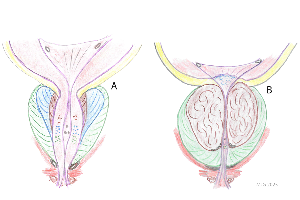
(B) Represents the aging man’s prostate with a moderately enlarged benign TZ narrowing the prostatic urethra and pushing the posterior CZ upward in the lower section of the bladder. It also partly compresses the PZ and indirectly the neuro-vascular network against the pelvic muscles. Calcifications appear around the urethra (black dots). The prostatic ducts are compressed and the direction altered. The drawings are inspired from private notes, the classical Sobotta and Becher anatomy textbook (München, 9th Engl edition 1975), the F Netter illustrations in the Ciba collection (1965) and McNeil’s description of the anatomical and histological zones [4].
4 The etymological meaning of prostatitis
The Greek physician and father of anatomy Herophilus of Chalcedon (around 335–255 BCE) is said to have been the first to describe the “Adenoides parastates” and even to have used the word “prostatês”, meaning literally “one who stands before”, [5]. Vesalius sketched the prostate in his anatomical drawings in 1543 [6], but the designation prostatae was first used in France around 1550–1600 by Dubois and Du Laurens. The latter mentioned “inflammation & ulcère des prostates” in “gonorrhoea Venerea” [7], and August Vidal was first to use in 1860 the word prostatitis for the same disease [8].
Prostatitis comes from the fusion of the word “prostatês” and the ending “-itis” expressing “inflammatio”, the Latin word for fire, glow and passion, a poetic metaphor well suitable for inflammation of the prostate.
5 Defining prostatitis in a global perspective
5.1 Present classification of clinical prostatitis
The present National Institute of Health (NIH) clinical classification of prostatitis dates to the 1990-ies, focusing on acute or chronic bacterial prostatitis (types I and II) and chronic non-bacterial prostatitis (type III) associated with a range of symptom combinations (prostatitis syndrome). In a fourth type (IV) – asymptomatic prostatitis – inflammatory components are detected in otherwise symptom free individuals [9]. In daily clinical praxis, prostatitis is predominantly referred to as chronic prostatitis (CP) type III in association with the chronic pelvic pain syndrome (CP/CPPS). However, inflammation of the prostate must be seen from both etymological and recent epidemiological and histopathological acquisitions in a notably broader meaning, and the health consequences needs to be viewed in a global demographic perspective.
5.2 Definition of prostatitis
Prostatitis is primarily a histopathological diagnosis proving mobilisation of the immune system including inflammatory cell infiltration (e.g. neutrophiles, lymphocytes, mast cells, others), presence of biomarkers (e.g. pro-inflammatory cytokines and interleukins (IL)), vascular dilatation and cellular damage [10]. These parameters are usually not available to the clinician to ascertain the diagnosis. Clinical definition is limited to symptoms and findings at clinical and laboratory examinations (Table 1A).
5.3 Prevalence of inflammatory infiltration
Inflammation is reported in a large number of prostate tissues removed by core biopsy, transurethral resection, and prostatectomy. In men over 60 years of age, a number of independently studies have identified various grades of inflammation in between 30 to 100% of the samples [10], [11], [12], [13], [14], [15], [16]. The inflammation follows the glandular tissue affecting ducts, tubules and acini leading to a disorganisation of their structure and infiltrates the stroma as well. In acute inflammation, Prostate Specific Antigen (PSA) leaks and raises often sharply in serum, as a sign of cellular disruption, taking months to return to nadir value [17]. Chronic inflammation affects less the PSA-value, showing relative stable quasi normal levels except in recurrent febrile infection [14], [17], [18].
The exact prevalence of inflammation in the prostate by age groups, especially in men under 60 years, is not well documented. The lack of an international consensus on the pathological classification is a limitation, although a few working schemes exist [16], [18]. The localisation in terms of zones and lobs is mostly lacking. Examples of inflammation are found in Attachment 1.
6 Diagnosis of prostatitis
The tools to define a state of inflammation in the prostate are limited. Clinical presentation varies widely and finding at digital rectal examination (DRE) is often ambiguous (Table 1A). The traditional 4-glass test may reveal an increased number of white blood cells in expressed prostatic secretion (EPS) indicative of inflammation and, inconsistently, bacterial growth in urine and EPS.
Imaging with transrectal ultrasound, computer tomography or magnetic resonance is not sufficiently characteristic to ascertain a diagnosis [19]. Pathological examination is the only true method. However, biopsy is ethically justifiable only for PCa detection and not for identifying inflammation or BPH, because of the high risk of a healthcare associated (HAI) febrile urogenital infection (UGI) [17]. Therefore, the finding of inflammation is mostly incidental and in an elderly population, but evidence is strong enough for giving it high importance.
Inflammatory biomarkers (e.g. IL-1, IL-4, Il-6, IL-8) of an inflamed prostate are expected to mirror those of urinary tract infections (UTI) [10] and have a great potential, but their quantification is limited by low values and low specificity [20].
7 The prostate-urine reflux loop
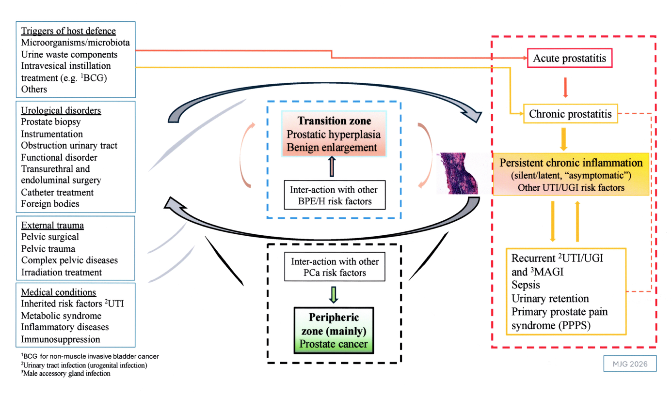
Reflux of urine into the prostatic ducts is well documented [21], [22]. There are reasons to believe that the urine and local epithelial secretion that reflux into the ducts and the prostatic fluid outflowing into the urethra are the carrier of microorganisms and other components. The elements identified in the prostatic fluid are therefore also interpreted as representative of the activity within the gland. The reflux mechanism in the aging prostate can further be alleged responsible for obstruction and stagnation in the excretory network leading to the formation of biofilms and calcifications, an environment favourable for harbouring microorganisms. Such mechanism is difficult to prove but it is reasonable to believe that the organ becomes more vulnerable (a locus minoris resistentiae), initiate recurrent infective events and possibly an inflammatory vicious cercle (Figure 1, Figure 2).
8 The triggers of inflammation
8.1 Host defence
In the urinary tract, the urinary flow “washes out” pathogens and the epithelial layer is the first barrier of defence. When pathogens attach to the epithelial layer on specific receptors – the Toll-like TLR4 receptors – the innate immune response is activated [23]. Sustained exposure to a “trigger” will lead to recruitment of lymphocytes and other components of the adaptive immune system turning the response to a chronic pattern. Little is known regarding the prostate, but there is reasonable evidence for a similar innate response [10].
8.2 The microbiome
The microbiome is nowadays recognised as the “community of microorganisms (bacteria, archaea, fungi, algae, viruses) and their genomes that live together within a reasonably well-defined habitat which has distinct physio-chemical properties” [24]. The microbiome consists of billions of microorganisms living on and inside the body in a homeostatic balance (eubiosis), stimulating the immune system in a way considered beneficial for health. There are different environments – “fields of activities” – such as the skin, the gut, the vagina, the urinary tract with “niche-specific” microbiota (all living members of a specific microbiome). The microbiome is dynamic in time and space and may vary with geographic regions, season, age, gender, diet, and reasonably also with travel, sexual partnership, antibiotic use, among others. Alterations in the composition or the physiology of the habitat (dysbiosis) may start a sequence of events impacting on the immune system and initiating disease [25], [26].
Uropathogens represent only a tiny fraction of the microbiota. The Gram-negative and some Gram-positive bacterial strains are the best established uropathogens invading the prostatic tissue (Table 1B). However, the impact of the microorganisms in the different zones of activity of the prostate is enigmatic.
8.3 Sexually transmitted infections
The knowledge about the role of STI-pathogens beside gonorrhoeal infection is limited and to some extend still controversial. Chlamydia trachomatis (C Trachomatis) and some viruses merit special attention.
8.3.1 Chlamydia trachomatis
According to the World Health Organization (www.who.int), C. trachomatis infection is the most frequent bacterial STI worldwide with over 128 million cases reported [27], probably only the top of the iceberg. In the European region, the prevalence of C. trachomatis detection in men is estimated to be at least 2.2% (range 2–6%, or more). Of these, 85% are between 15–29 years of age and well above 50% are symptom free [28]. The natural clearance time after infection varies widely but reported at 2.84 years (= infection time!) for men [29]. In males, C. trachomatis is detected in the urethra, rectum, epididymis, buccal cavity and conjunctiva but its role in maintaining stimulation of the immune system in the prostate is insufficiently investigated. Recurrence or re-infection is not well documented except in men having sex with men [30]. Studies have also shown multiple microorganisms in men of all ages presenting with male accessory gland infections (MAGI) [31].
8.3.2 Viruses
Human papillomavirus (HPV) is probably the most common of all STI worldwide, often found in co-infection with C. trachomatis [32], [33]. Many HPV-types are known to cause diseases in humans (e.g. types 6, 11, 16, 18) including condyloma, cancer of the anus, the oropharynx, and the penis [33]. A meta-analysis showed HPV in 26% of PCa tissue, 17% in BPH/E and 9% of unaltered prostate tissue and therefore considered as a risk factor for PCa [34]. In women a few types of HPV (type 16, 18) are high-risk associated with cervical cancer and vaccination of young women aims at preventing and even eradicating cervical cancer [35]. Vaccination of young boys is also recommended by the WHO (www.who.int). Vaccination reporting is missing in many countries, and available rates vary widely from none or negligeable to over 70% in some European countries (e.g. UK, Sweden; gov official sites, Apr 2026).
Against this background, understanding 1) the role of latent cancerogenic viruses harbored in the prostate, and 2) the semen as carrier of STI viruses is of utmost importance [36].
8.4 Other factors impacting prostate pathophysiology
Besides infectious factors stimulating the host defense there are multiple medical, surgical and traumatic risk factors that directly or indirectly act on the prostate. Surgical instrumentation, especially transurethral and catheterization, directly affect the uroepithelial layer around the ductal outlets. Lower urinary tract dysfunction perturbs the urinary flow. Complex pelvic surgery, targeted irradiation and other traumatic events disturb the physiological functions in the pelvic area (Figure 2).
9 The role of animal models
Due to the difficulties in studying activities within the human prostate, in-vitro and in-vivo experiments are important complement to our understanding of series of events occurring in the prostate. Intraprostatic γ-carrageenan injection induces prostate inflammation and pain [10], [37], [38]. Studies have shown that a few specified medical products have anti-inflammatory effects and reduce nociceptive pain, curb cell proliferation, and expression of androgen receptors and biomarkers [38]. A difficulty resides in the translation of finding in animals to diseases in humans.
10 Discussion on inflammation and the three prostate pathologies
10.1 Benign prostatic enlargement
Benign hyperplasia and nodular formation are principally seen in the TZ (Figure 1B) and regarded as a normal process of aging, starting already around the age of 40 and affecting 80% of men over 70 [2], [39], [40]. BPH/E affects quality of life through lower urinary tract symptoms (LUTS) and increased risk of UTI, followed by disabilities through neuro-muscular bladder dysfunction, urinary retention, and finally of upper urinary tract obstruction, renal failure and death.
The global burden of disease 2021 report states a prevalence of BPH/E over 112.5 million with an estimated increase of 122% since 1990 and a markedly expected increase the coming decades [41]. In men over 60 years, some grade of inflammation is reported in more than 70% of all BPH/E specimens [11], [13], [14], [15], [39]. The role of the microbiome as an inducer is given priority concern [42], [43], [44], and the calcifications and biofilms, mostly located dorsally between the TZ and PZ, are suitable anchorage of latent, undetectable, microorganisms.
Age is not a risk factor by itself, but advanced age is rather the period of life risk factors act and interact. These are essentially a) age-related programmed decrease in androgen production; b) heredity (families with BPH/E); c) inflammation and d) immunological disfunction; e) lifestyle (e.g. smoking, diet, alcohol consumption, reduced physical activity) and associated f) metabolic syndrome; g) vascular insufficiency and hypoxia. Their inter-interaction is poorly understood [2], [40], [45], [46], [47], [48], [49].
10.2 Prostate cancer
PCa is worldwide the second most commonly cancer and the fifth leading cause of cancer death among men, with over 1,460,000 estimated cases and 396,000 deaths in 2022, with a marked geographic variation [50], [51]. Most PCa are located in the PZ and inflammation of various extent and grade of severity is found in the micro-environment in over 50% of the samples [4], [11], [12], [13], [52], [53], [54], [55]. Most authors agree that the human microbiome can maintain a chronic inflammation, generate cellular alterations and “proliferative inflammatory atrophy”, followed by “prostatic intraepithelial neoplasia”, a predecessor of PCa [52], [53], [54], [55], [56], [57]. As for BPH, a key issue is how inflammation interacts with the other risk factors such as heredity, immunity, diet and lifestyle, and toxic carcinogenetic elements present in our environment.
10.3 Clinical prostatitis complex
Table 1 (Table 1B) gives an overview of the infectious and inflammatory prostatitis.
The spontaneous invasion route in acute bacterial prostatitis is believed to be retrograde through the ducts although some may be blood-born. The histopathological finding is believed to be that of an acute “stormy” host response, as in febrile systemic UTI (type I-A) [23]. However, hypothetically, a mild form is plausible in clinical ambiguous situations or symptom free infections that eventually could convert to persisting inflammation (type I-B). A few studies indicate that up to 10% of acute prostatitis may convert into chronic infections [58].
Chronic bacterial prostatitis (type II-A) is a microbiological proven colonisation of the prostate (bacterial growth in urine and EPS), with the potential of recurrent episodes. The histopathology is assumed to be that of chronic profile, pinpointing the role of inflammation as a harbour of latent microorganisms that precisely initiated the immune response.
Mycobacterium tuberculosis can invade the prostate and once established on site will induce a chronic granulomatous prostatitis (type II-C) and purulent secretion in urine and EPS. The urogenital tracts (kidney, ureter, bladder, genital glands) are the two most important extrapulmonary sites of tuberculous infections in endemic region and often associated with HIV [59]. Other forms of non-tuberculous granulomatous prostatitis are also mentioned in Table 1B (type III-C [60].
Symptom-associated CP without proven bacterial involvement (type III) is the largest symptomatic group and challenging for medical management. A large number of independent studies and population-bases investigations have revealed that about 10% (range 3–14%) of all men are affected [61], [62]. The finding of inflammatory cells in prostatic secrete (Type IIIA) is a rough indicator of what might happen within the prostate, and, ironically, the evidence of any histological proven inflammatory process is weak.
The fourth group (type IV) is nowadays understood as a group of men experiencing no or ill-defined symptoms but presenting an excess of leucocytes (pus) in the EPS, a slight PSA elevation or a histologically proven inflammation of the prostate alone usually in association with BPH/E or PCa. Noteworthy, the expression “asymptomatic” is relative as with age, individuals’ own empirical perception of mild to moderate LUTS or discomfort is often accepted as within “normal” range [63]. This group is obviously now to be considered as the overall largest one with demonstrated links to BPH/E and PCa (Type IV-A, B) (Tables 1A, 1B).
|
Infection type/Genesis |
Occurrence |
Clinical finding |
|
Type I. Acute prostatitis, microorganism1 associated – identified or expected (urine, blood samples) |
||
|
A. Acute febrile, urogenital pathogens associated |
Spontaneous infection uncommon. |
Range from slight fever, LUTS2 and local pain to life-threatening systemic infection/urosepsis. Risk of residual urine and urinary retention. Rarely develops prostatic abscess. DRE3: inflamed, tenderness |
|
B. Asymptomatic or presumed low-grade, insidious infection microorganisms associated (e.g. STI) |
Gonorrhoea well documented, increasing globally. Role ill-defined for other organisms. Recurrence or re-infection may occur |
From sub-clinical to cystitis or urethritis-like infection, epididymis, prostatitis. Untreated gonorrhoea can be destructive for the prostate (e.g. abscess). DRE3: no, limited, variable finding as above |
|
Type II. Chronic prostatitis, microorganism associated – identified or expected (urine, EPS4) |
||
|
A. Persistence of urogenital pathogens |
Prevalence not well documented. High risk of recurrent infectious episodes |
Variable LUTS2 and local pelvic pain. Switch between asymptomatic and symptomatic episodes. Risk of evolving into primary prostate pain syndrome (PPPS). DRE3: variable, poor correlation with symptoms |
|
B. Associated with other microorganisms (e.g. STI) |
Controversial and role ill-defined. Proven case reports/series. Risk of recurrence |
From asymptomatic to variable LUTS2 and pelvic and regional pain. Risk of evolving into PPPS |
|
C. Urogenital tuberculosis |
Overall uncommon. Prevails in tuberculosis high endemic geographic areas, countries. Most common extra-pulmonary tuberculosis. Risk associated with HIV- infection |
Occurs in kidney, ureter, urinary bladder, urethra, epididymis, prostate. Risk of scrotal and perineal fistula development. DRE3: variable. Can mimic prostate cancer |
|
D. Urogenital Schistosomiasis (lower urinary tract and male accessory glands) |
Mostly uncommon except in endemic areas (water, rivers, lakes infected with intermediate host – snails) |
Haematuria, sandy patches in bladder. Scars and strictures in ureter, urinary bladder, urethra. Risk factor of urinary bladder cancer |
|
Type III. Chronic prostatitis – no identified or proven microorganism. Prostatitis syndrome |
||
|
A. Presence of leucocytes in EPS4 |
Affect 3–10% of male population. Important geographic variations |
Wide range of symptoms such as LUTS2, regional and pelvic pain, functional and sexual dysfunction, anxiety. Phenotyping and identification of symptom profile using UPOINT5 |
|
B. Absence of leucocytes in EPS |
||
|
C. Granulomatous prostatitis |
Uncommon |
Variable LUTS2, dysuria, haematuria, febrility. Often associated with co-morbidity. DRE3: mimic prostate cancer |
|
Type IV. Proven inflammatory infiltration without identified microorganism or typical prostate-related symptoms (also called asymptomatic prostatitis) |
||
|
A. Age related process |
High prevalence. Slight PSA elevation observed (variable)
|
No symptoms reported. A relative term considering patients’ perception of “normality” [63] |
|
B. Disease associated (sub-groups according to link to different medical conditions) |
a) BPH/E b) PCa c) Metabolic syndrome d) Other inflammatory diseases e) Trauma, pelvic surgery, irradiation |
Related to underlying cause and co-morbidity. Metabolic syndrome with impact on inflammation. DRE: variable, cause related |
1Microorganisms according to the National Cancer Institute/National Institute of Health: include bacteria, protozoa, algae, fungi and virus
2LUTS: Lower urinary tract symptoms
3DRE: Digital rectal examination
4EPS: Expressed prostatic secretion
5UPOINTS: Phenotypes according to dominating symptom profile or domain: Urinary, Psychosocial, Organ specific, Infectious, Neurological, Tenderness, sexual dysfunction
|
Infection type/Genesis |
Microbiology |
Histopathology |
|
Type I. Acute prostatitis, microorganism1 associated – identified or expected (urine, blood samples) |
||
|
A. Acute febrile, urogenital pathogens associated |
Escherichia coli Enterobacterial species Klebsiella species Pseudomonas species Enterococcus species Growth in urine and/or blood |
Acute inflammation pattern. See Attachment 1 |
|
B. Asymptomatic or presumed low-grade, insidious infection microorganisms associated (e.g. STI) |
Neisseria gonorrhoea Chlamydia trachomatis Viruses (e.g. Human Papillomavirus HPV) Other microorganisms poorly investigated. Growth in urine and EPS4 |
Histopathology usually unknown except for Gonorrhoeal infection. |
|
Type II. Chronic prostatitis, microorganism associated – identified or expected (urine, EPS4) |
||
|
A. Persistence of urogenital pathogens |
Gram-negative species as acute infections. Gram-positive species. Candida infection reported. New detected species to be confirmed. Growth in urine and EPS4 |
Mixed acute and chronic inflammatory pattern expected but mostly not proven |
|
B. Associated with other microorganisms (e.g. STI) |
Chlamydia trachomatis Ureaplasma urealyticum Viruses (e.g. HPV, other viruses) |
|
|
C. Urogenital tuberculosis |
Mycobacterium tuberculosis. ZN6-coloration for identification in urine and prostatic tissue |
Chronic specific granulomatous inflammation: caseous necrosis, epithelial granuloma and giant cells. |
|
D. Urogenital Schistosomiasis (lower urinary tract and male accessory glands) |
Schistosoma haematobium Schistosoma eggs found in urine |
Chronic granulomatous inflammation with fibrosis |
|
Type III. Chronic prostatitis – no identified or proven microorganism. Prostatitis syndrome |
||
|
A. Presence of leucocytes in EPS4 |
No microorganisms identified with routine laboratory methods. New strains of the urogenital microbiome under investigation |
Chronic inflammation proven or presumed |
|
B. Absence of leucocytes in EPS |
||
|
C. Granulomatous prostatitis |
Unspecified |
Chronic granulomatous inflammation. Characteristic Xanthogranulomatous pattern in very rare cases |
|
Type IV. Proven inflammatory infiltration without identified microorganism or typical prostate-related symptoms (also called asymptomatic prostatitis) |
||
|
A. Age related process |
No pathogens identified. Microorganisms from the urogenital microbiome under investigation (e.g. HPV, others) |
Inflammatory infiltration found in both glandular and stromal tissues (predominantly chromic pattern, remodelling of prostate gland. Incidental finding |
|
B. Disease associated (sub-groups according to link to different medical conditions) |
||
1Microorganisms according to the National Cancer Institute/National Institute of Health: include bacteria, protozoa, algae, fungi and virus
4EPS: Expressed prostatic secretion
6ZN-coloration = specific Ziel-Nielsen specific coloration for microscopic identification of Mycobacterium tuberculosis
10.4 Men’s urogenital infections
It is often challenging to differentiate between the urinary tract and the prostate as primary point of departure of a symptomatic infection [64]. PSA often increases notably in febrile UTI and systemic infections, indicating involvement of the prostate [14], [17]. The term urogenital infection (UGI) might therefore be preferred in men, especially in case of febrile infection. Their number in a global perspective is difficult to estimate. Including all forms of non-febrile UTI (e.g. cystitis) and UGI the number must be enormous - innumerable huge. Considering the easiness of access to antimicrobials on prescription as well as “over the counter”, the antibiotic treatment episodes must reach gigantesque quantities and contribute directly to the antimicrobial resistance development and selection of multi-resistant microorganisms in the environment.
Sepsis is recognised as a global health issue reported in over 49 million cases and 11 million deaths in 2020 [65]. Several studies have pointed out the urogenital tracts to be the origin in 20–25% of all sepsis cases, with a reported mortality rate ranging from a few to over 20% depending on age, comorbidity, severity of sepsis, access to health care, timing of antibiotic treatment and bacterial species. In a recent Swedish targeted study, the mortality rate was 14% [66]. With regard to the uncertainty in the reporting of sepsis and sever UGI globally, it would not be disrespectful to consider the data as an underestimate of the true reality.
11 Prospectives
The quintessence of this viewpoint is the enormous global impact on men’s health of the three prostatic diseases together. There is beyond any doubts that inflammation, caused by a succession of infections and other traumatic impacts, is a key risk factor. A reasonable hypothesis is that the development of the three diseases results from a lifelong process debuting in the young adult, boosted at some point in the early midlife by a constellation of pathophysiological events culminating above 70 years of age. The inflammatory process appears to interact with the other disease-specific risk factors differently in the different anatomical zones (Figure 2). To better understand the development, the urological community needs to update and improve knowledge in several sectors and for that needs increased research resources.
11.1 Epidemiology and classifications
A validated histopathological classification of inflammation of the prostate should be established and the identified pattern in each case reported by the pathologist (i.e. acute versus chronic, extent, distribution, zone). An update of the current classification of prostatitis should reflect the global and broader meaning of prostatitis to strengthen diagnosis reporting, teaching and research. A tentative upgrade is given in (Tables 1A, 1B) including a larger number of causative agents (e.g. tuberculous, parasites, STI, viruses) as potential initiator of the immune host response creating an intraprostatic inflammation. The tables are amended to be a pragmatic and tutorial tool reflecting the occurrence, clinical manifestation, known microbiologic findings and proven or expected histopathology within the different groups. The prevalence of intraprostatic inflammation by age groups (even younger when available) should be studied.
11.2 Diagnostic tools
As for PCa, a low invasive imaging tool is highly desirable, to be used in combination with improved knowledge about inflammatory biomarkers.
11.3 Microbiology
Research should focus on defining the microorganisms of the urogenital microbiome capable of maintaining inflammation and the way this succession of events interacts with the incriminated disease-specific risk factors in the prostate’s different zones [67], [68].
11.4 Metabolic syndrome
Progress in the understanding of the complex bidirectional interaction between inflammation and the metabolic syndrome in men and how it putatively also affects the prostate.
11.5 Access to health care and prevention
Only a minority of the aging male population worldwide will be spared from the burden of one or more of the prostatic diseases. Keeping in mind that about half of the world population (mainly in low-income countries) do not have access to essential health and surgical care [69], a challenging issue is whether affordable medical control (including targeted vaccination) of the inflammatory process could reduce or delay BPH/E and PCa development, curb the load of symptomatic infections and antibiotic use and alleviate the burden of the prostatitis syndrome. The objectives are clear: lessen the patients’ suffering and reduce costs. Additional research funding is necessary to encourage knowledge in these fields, improve teaching and research, and promote prevention against prostatic diseases.
Abbreviations
| BCE | Before the common era (= also BC) |
| BPH/E | Benign prostatic hyperplasia/enlargement |
| CP/CPPS | Chronic prostatitis/Chronic pelvic pain syndrome |
| C trachomatis | Chlamydia trachomatis |
| CZ | Central zone (of the prostate) |
| DRE | Digital rectal examination |
| EPS | Expressed prostatic secretion |
| HAI | Health care associated infection |
| IL | Interleukins |
| LUTS | Lower urinary tract symptoms |
| MAGI | Male accessory gland infection |
| PCa | Prostate carcinoma |
| PPPS | Primary prostate pelvic syndrome |
| PZ | Peripheral zone of the prostate |
| STI | Sexually transmitted infection |
| TZ | Transition zone of the prostate |
| UGI | Urogenital infection |
| UPOINT | Phenotyping of prostatic syndromes (see Table 1, footnote) |
| UTI | Urinary tract infection |
| ZN | Ziel-Nielsen staining for microscopy of Mycobacterium tuberculosis |
Author’s ORCID
Magnus J Grabe: 0000-0002-4449-9048
Competing interest
None
Funding
None. A private initiative without any external funding
References
[1] Lowsley OS. The development of the human prostate gland with references to the development of other structures of the neck of the urinary bladder. Am J Anat. 1912;13:299-349. DOI: 10.1002/aja.1000130303[2] Aaron L, Franco OE, Hayward SW. Review of prostate anatomy and embryology and the etiology of Benign Prostatic Hyperplasia. Urol Clin North Am. 2016;43:279-88. DOI: 10.1016/j.ucl.2016.04.012
[3] Bucher O. Cytologie, Histologie und Mikroskopische Anatomie des Menschen. 5th edition. Bern: Hans Huber Verlag; 1967.
[4] McNeal JE. The zonal anatomy of the prostate. Prostate. 1981;2:35-49. DOI: 10.1002/pros.2990020105
[5] Von Staden H. Herophilus. The art of medicine in early Alexandria. Cambridge: Cambridge University press; 1989.
[6] Vesalius A. De humani corporis fabrica libri septem. Ex officina Joannis Oporini; 1543. Liber V, plate 59.
[7] Du Laurens A. L’Histoire anatomique en laquelle toutes les parties du corps hulain sont amplement declarees enrichie de controverses et observations nouvelles...de la traduction de Francois Size [Historia anatomica humani corporis et singularum eius partium multis controuersis & obseruationibus nouis illustrata]. Paris; 1610. Available from: https://www.biusante.parisdescartes.fr/histmed/medica/cote?31655
[8] Vidal A. Traité de pathologie externe et de médecine opératoire Tome 4. Paris, New York: J-B Baillière; 1860. p. 721.
[9] Krieger JN, Lee SW, Jeon J, Cheah PY, Liong ML, Riley DE. Epidemiology of prostatitis. Int J Antimicrob Agents. 2008 Feb;31 Suppl 1(Suppl 1):S85-90. DOI: 10.1016/j.ijantimicag.2007.08.028
[10] Dong-Ru Ho. Prostate inflammation: A brief review. Urol Sci. 2017;28(3):113-118. DOI: 10.1016/j.urols.2017.04.003
[11] Yli-Hemminki TH, Laurila M, Auvinen A, Määttänen L, Huhtala H, Tammela TL, et al. Histological inflammation and risk of subsequent prostate cancer among men with initially elevated serum prostate-specific antigen (PSA) concentration in the Finnish prostate cancer screening trial. BJU Int. 2013 Oct;112(6):735-41. DOI: 10.1111/bju.12153
[12] Engelhardt PF, Brustmann H, Seklehner S, Riedl CR. Chronic asymptomatic inflammation of the prostate type IV and carcinoma of the prostate: is there a correlation? Scan J Urol. 2013;47:230-235. DOI: 10.3109/00365599.2012.733961
[13] Edlin RS, Heyns CF, Van Vuuren SP, Zarrabi AD. Prevalence of histological prostatitis in men with benign prostatic hyperplasia or adenocarcinoma of the prostate presenting without urinary retention. S Afr J Surg. 2012 Nov 12;50(4):127-30. DOI: 10.7196/sajs.1095
[14] Schatteman PH, Hoekx L, Wyndaele JJ, Jeuris W, Van Marck E. Inflammation in prostate biopsies of men without prostatic malignancy or clinical prostatitis: correlation with total serum PSA and PSA density. Eur Urol. 2000 Apr;37(4):404-12. DOI: 10.1159/000020161
[15] Zlotta AR, Egawa S, Pushkar D, Govorov A, Kimura T, Kido M, et al. Prevalence of inflammation and benign prostatic hyperplasia on autopsy in Asian and Caucasian men. Eur Urol. 2014 Oct;66(4):619-22. DOI: 10.1016/j.eururo.2014.06.026
[16] Allott EH, Markt SC, Howard LE, Vidal AC, Moreira DM, Castro-Santamaria R, et al. Geographic differences in baseline prostate inflammation and relationship with subsequent prostate cancer risk: results from the multinational REDUCE trial. Cancer Epidemiol Biomarkers Prev. 2018 Jul;27(7):783-789. DOI: 10.1158/1055-9965.EPI-18-0076
[17] Ulleryd P, Sandberg T. Ciprofloxacin for 2 or 4 weeks in the treatment of febrile urinary tract infection in men: a randomized trial with a 1 year follow-up. Scand J Infect Dis. 2003;35(1):34-9. DOI: 10.1080/0036554021000026988
[18] Irani J, Levillain P, Goujon JM, Bon D, Doré B, Aubert J. Inflammation in benign prostatic hyperplasia: correlation with prostate specific antigen value. J Urol. 1997 Apr;157(4):1301-3. DOI: 10.1016/s0022-5347(01)64957-7
[19] Kitzing YX, Prando A, Varol C, Karczmar GS, Maclean F, Oto A. Benign conditions that mimic prostate carcinoma: MR imaging features with histopathologic correlation. Radiographics. 2016 Jan-Feb;36(1):162-75. DOI: 10.1148/rg.2016150030
[20] Horváth J, Wullt B, Naber KG, Köves B. Biomarkers in urinary tract infections - which ones are suitable for diagnostics and follow-up? GMS Infect Dis. 2020 Nov 26;8:Doc24. DOI: 10.3205/id000068
[21] Blacklock NJ. Anatomical factors in prostatitis. Br J Urol. 1974 Feb;46(1):47-54. DOI: 10.1111/j.1464-410x.1974.tb10158.x
[22] Kirby RS, Lowe D, Bultitude MI, Shuttleworth KE. Intra-prostatic urinary reflux: an aetiological factor in abacterial prostatitis. Br J Urol. 1982 Dec;54(6):729-31. DOI: 10.1111/j.1464-410x.1982.tb13635.x
[23] Ambite I, Butler D, Wan MLY, Rosenblad T, Tran TH, Chao SM, Svanborg C. Molecular determinants of disease severity in urinary tract infection. Nat Rev Urol. 2021 Aug;18(8):468-486. DOI: 10.1038/s41585-021-00477-x
[24] Berg G, Rybakova D, Fischer D, Cernava T, Vergès MC, Charles T, et al. Microbiome definition re-visited: old concepts and new challenges. Microbiome. 2020 Jun 30;8(1):103. DOI: 10.1186/s40168-020-00875-0
[25] Manos J. The human microbiome in disease and pathology. APMIS. 2022 Dec;130(12):690-705. DOI: 10.1111/apm.13225
[26] Fernandez E, Wargo JA, Helmink BA. The Microbiome and cancer: a translational science review. JAMA. 2025 Jun 24;333(24):2188-2196. DOI: 10.1001/jama.2025.2191
[27] Mohseni M, Sung S, Rausch-Phung EA, et al. Chlamydia. [Updated 2026 Jan 10]. In: StatPearls [Internet]. Treasure Island (FL): StatPearls Publishing; 2026 Jan-. Available from: https://www.ncbi.nlm.nih.gov/books/NBK537286/
[28] Velicko I. Chlamydia trachomatis infection in Sweden: time trends, risk factors, and prevalence [Thesis]. Stockholm, Sweden: Karolinska Institutet; 2021.
[29] Lewis J, Price MJ, Horner PJ, White PJ. Genital Chlamydia trachomatis Infections clear more slowly in men than women, but are less likely to become established. J Infect Dis. 2017 Jul 15;216(2):237-244. DOI: 10.1093/infdis/jix283
[30] Williams E, Williamson DA, Hocking JS. Frequent screening for asymptomatic chlamydia and gonorrhoea infections in men who have sex with men: time to re-evaluate? Lancet Infect Dis. 2023 Dec;23(12):e558-e566. DOI: 10.1016/S1473-3099(23)00356-0
[31] Pilatz A, Hossain H, Kaiser R, Mankertz A, Schüttler CG, Domann E, et al. Acute epididymitis revisited: impact of molecular diagnostics on etiology and contemporary guidelines recommendations. Eur Urol. 2015;68:428-35. DOI: 10.1016/j.eururo.2014.12.005
[32] Toh ZQ, Zhao F, Zhang L, Wei L, Zou H. Editorial: HPV natural history, immunological responses and vaccination strategies: challenges and opportunities. Front Immunol. 2024 Jan 8;14:1349664. DOI: 10.3389/fimmu.2023.1349664
[33] Sarier M, Ceyhan AM, Sepin N, Ozel E, Inal MM, Kukul E, et al. HPV infection in urology practice. Int Urol Nephrol. 2020;52:1-8. DOI: 10.1007/s11255-019-02302-2
[34] Tsydenova IA, Ibragimova MK, Tsyganov MM, Litviakov NV. Human papillomavirus and prostate cancer: systematic review and meta-analysis. Sci Rep. 2023;13(1): 16597. DOI: 10.1038/s41598-023-43767-7
[35] Choi S, Ismail A, Pappas-Gogos G, Boussios S. HPV and Cervical cancer: a review of epidemiology and screening uptake in the UK. Pathogens. 2023;12:298. DOI: 10.3390/pathogens12020298
[36] Sucato A, Buttà M, Bosco L, Di Gregorio L, Perino A, Capra G. Human Papillomavirus and male infertility: what do we know? Int J Mol Sc. 2023;24:17562. DOI: 10.3390/ijms242417562
[37] Zeng F, Chen H, Yang J, Wang L, Cui Y, Guan X, et al. Development and validation of an animal model of prostate inflammation-induced chronic pelvic pain: evaluating from inflammation of the prostate to pain behavioral modifications. PloS One. 2014;9(5):e96824. DOI: 10.1371/journal.pone.0096824
[38] Chabot S, Dizeyi N, Ramnemark L, Lluel P, Abrahamsson P-A, Grabe M. Impact of CernitinTM on induced chronic prostatitis in animal model for understanding management of lower urinary tract symptoms. Phytomed Plus. 2021;1(4):100057. DOI: 10.1016/j.phyplu.2021.100057
[39] Oesterling JE. The origin and development of benign prostatic hyperplasia An age-dependent process. J Androl. 1991;12:348-355. DOI: 10.1002/j.1939-4640.1991.tb00271.x
[40] Berry SJ, Coffey DS, Walsh PC, Ewing LL. The development of human benign prostatic hyperplasia with age. J Urol. 1984;132(3):474-9. DOI: 10.1016/S0022-5347(17)49698-4
[41] Xin Chen, Siyuan Y, Zhangxin He, Chen Z, Tang X, Lin Y, et al. Comprehensive analysis of the global, regional, and national burden of benign prostatic hyperplasia from 1990 to 2021. Scientific Reports. 2025;15:5644. DOI: 10.1038/s41598-025-90229-3
[42] Perez-Carrasco V, Soriano-Lerma A, Soriano M, Gutiérrez-Fernández J, Garcia-Salcedo JA. Urinary microbiome: Yin and Yang of the urinary tract. Front Cell Infect Microbiol. 2021;11:617002. DOI: 10.3389/fcimb.2021.617002
[43] Dalvi H, De Nisco NJ. The evolving world of the urinary microbiome. Curr Opin Urol. 2024 Nov 1;34(6):422-427. DOI: 10.1097/MOU.0000000000001222
[44] Gilbert NM, Ackerman AL, Lewis AL. Editorial: The urogenital microbiota in urinary tract diseases. Front Cell Infect Microbiol. 2022 Dec 8;12:1096168. DOI: 10.3389/fcimb.2022.1096168
[45] Devlin CM, Simms MS, Maitland NJ. Benign prostatic hyperplasia - what do we know? BJU Int. 2021 Apr;127(4):389-399. DOI: 10.1111/bju.15229
[46] Cannarella R, Condorelli RA, Barbagallo F, La Vignera S, Calogero AE. Endocrinology of the aging prostate: current concepts. Front Endocrinol (Lausanne). 2021 Feb 22;12:554078. DOI: 10.3389/fendo.2021.554078
[47] Xu G, Dai G, Huang Z, Guan Q, Du C, Xu X. The Etiology and pathogenesis of benign prostatic hyperplasia: the roles of sex hormones and anatomy. Res Rep Urol. 2024 Sep 23;16:205-214. DOI: 10.2147/RRU.S477396
[48] Penna G, Fibbi B, Amuchastegui S, Cossetti C, Aquilano F, Laverny G, et al. Human benign prostatic hyperplasia stromal cells as inducers and targets of chronic immuno-mediated inflammation. J Immunol. 2009 Apr 1;182(7):4056-64. DOI: 10.4049/jimmunol.0801875
[49] Kramer G, Mitteregger D, Marberger M. Is benign prostatic hyperplasia (BPH) an immune inflammatory disease? Eur Urol. 2007 May;51(5):1202-16. DOI: 10.1016/j.eururo.2006.12.011
[50] Zhang W, Cao G, Wu F, Wang Y, Liu Z, Hu H, et al. Global Burden of prostate cancer and association with socioeconomic status, 1990-2019: a systemic analysis from the global burden of disease study. J Epidemiol Glob Health. 2023 Sep;13(3):407-421. DOI: 10.1007/s44197-023-00103-6
[51] Schafer EJ, Laversanne M, Sung H, Soerjomataram I, Briganti A, Dahut F, et al. Recent patterns and trends in global prostate cancer incidence and mortality: an update. Eur Urol. 2025;87(3):302-313. DOI: 10.1016/j.eururo.2024.11.013
[52] Sfanos KS, Yegnasubramanian S, Nelson WG, De Marzo AM. The inflammatory microenvironment and microbiome in prostate cancer development. Nat Rev Urol. 2018;15(1):11-24. DOI: 10.1038/nrurol.2017.167
[53] Liss MA, White JR, Goros M, Gelond J, Leach R, Johnson-Pais T, et al. Metabolic biosynthesis pathways identified from fecal microbiome associated with prostate cancer. Eur Urol. 2018;74(5):575-82. DOI: 10.1016/j.eururo.2018.06.033
[54] Markoswski MC, Boorjian SA, Burton JP, Burton JP, Hahn NM, Ingersoll MA, et al. The microbiome and genitourinary cancer: A collaborative review. Eur Urol. 2019;75(4):637-646. DOI: 10.1016/j.eururo.2018.12.043
[55] Fujita K, Matsushita M, De Velasco MA, Hatano K, Minami T, Nonomura N, et al. The gut-prostate axis: a new perspective of prostate cancer biology through the gut microbiome. Cancers (Basel). 2023 Feb 21;15(5):1375. DOI: 10.3390/cancers15051375
[56] Hurst R, Meader E, Gihawi A, Rallapalli G, Clark J, Kay GL, et al. Microbiome of urine and the prostate are linked to human prostate cancer risk groups. Eur Urol Oncology. 2022;5(4):412-19. DOI: 10.1016/j.euo.2022.03.006
[57] De Bono JS, Guo C, Gurel B, De Marzo AM, Sfanos KS, Mani R, et al. Prostate carcinogenesis: inflammatory storms. Nat Rev Cancer. 2020;20(8):455-469. DOI: 10.1038/s41568-020-0267-9
[58] Yoon B-I, Kim S, Han DS, Ha US, Lee SJ, Kim HW, et al. Acute bacterial prostatitis: how to prevent and manage chronic infection? J Infect Chemother. 2012;18(4):444-50. DOI: 10.1007/s10156-011-0350-y
[59] Kulchavenya E, Yasuda M, Grabe M. Epidemiology, classification, clinical features, and diagnostics of urogenital tuberculosis. Version: 2019-05-28. In: Bjerklund Johansen TE, Wagenlehner FME, Matsumoto T, Cho YH, Krieger JN, Shoskes D, Naber KG, editors. Urogenital Infections and Inflammations. Berlin: German Medical Science GMS Publishing House; 2017-. DOI: 10.5680/lhuii000052
[60] Kumbar R, Dravid N, Nikumbh D, Patil A, Nagappa KG. Clinicopathological overview of granulomatous prostatitis: an appraisal. J Clin Diagn Res. 2016;10(1):EC20-3. DOI: 10.7860/JCDR/2016/15365.7146
[61] Magistro G, Wagenlehner FME, Grabe M, Weidner W, Stief CG, Nickel JC. Contemporary management of chronic prostatitis/chronic pelvic pain syndrome. Eur Urol. 2016;69(2):286-97. DOI: 10.1016/j.eururo.2015.08.061
[62] Engeler D, Messelink EJ, Almeida Pinto R, Baranowski AP, Berghmans LCM, Birch J, et al. EAU guidelines on cronic pelvic pain. Edn. presented at EAU Congress, Madrid 2025. Available from: https://www.uroweb.org/guidelines
[63] Malde S, Umbach R, Wheeler J, Lytvyn L, Cornu JN, Gacci M, et al. A systematic review of patients' values, preferences, and expectations for the diagnosis and treatment of male lower urinary tract symptoms. Eur Urol. 2021;79(6):796-809. DOI: 10.1016/j.eururo.2020.12.019
[64] Kranz J, Bartoletti R, Bruyère F, Cai T, Geerlings S, Köves B, et al. European Association of Urology guidelines on urological infections: summary of the 2024 guidelines. Eur Urol. 2024;86(1):27-41. DOI: 10.1016/j.eururo.2024.03.035
[65] World Health Organisation. Fact sheet: Sepsis: Key facts. Geneva: World Health Organization; 2026. Available from: https://www.who.int/en/news-room/fact-sheets/detail/sepsis
[66] Holmbom M, Andersson M, Grabe M, Peeker R, Saudi A, Styrke J, et al. Community-onset urosepsis: incidence and risk factors for 30-day mortality - a retrospective cohort study. Scan J Urol. 2022;56(5-6):414-420. DOI: 10.1080/21681805.2022.2123039
[67] Oseni SO, Naar C, Pavlovic M, Asghar W, Hartmann JX, Fields GB, et al. The molecular basis and clinical consequences of chronic inflammation in prostatic diseases: Prostatitis, Benign Prostatic Hyperplasia, and Prostate Cancer. Cancers. 2023;15(12):3110. DOI: 10.3390/cancers15123110
[68] Wang Y, Chen G, Li D, Zhang D, Xing Q. Prostatitis, benign prostatic hyperplasia, and prostate cancer: a bidirectional Mendelian randomization study and clinical implications for these patients' populations. Biol Direct. 2024;19(1):129. DOI: 10.1186/s13062-024-00575-x
[69] Nepogodiev D, Picciochi M, Ademuyiwa A, Adisa A, Agbeka AE, Aguilera ML, et al. Surgical health policy 2025-35: strengthening essential services for tomorrow's needs. Lancet. 2025 Aug 23;406(10505):860-880. DOI: 10.1016/S0140-6736(25)00985-7
Attachments
| Attachment 1 | lhuii000076_att1 |
| Type | application/pdf |
| MD5 | 475723cfc002303564665e905a762d4d |
| SHA256 | e8cb5a5bd39560bc675ef3f1f81680037701389d5da895a585e15138b060a73c |