
[Laborbasierte Überwachung der Carbapenem-Resistenz bei klinischen Enterobacterales-Stämmen in einem nigerianischen Tertiärkrankenhaus: Nachweis der blaNDM- und blaVIM-Gene]
Abubakar Abubakar 1Garba Lawal 2
Idris Nasir Abdullahi 3
1 Gombe State Hospital Services Management Board, Gombe, Nigeria
2 Department of Microbiology, Faculty of Science, Gombe State University, Gombe, Nigeria
3 Department of Medical Laboratory Science, Faculty of Allied Health Sciences, College of Medical Sciences, Ahmadu Bello University, Zaria, Nigeria
Zusammenfassung
Hintergrund: Carbapenem-resistente Enterobacterales (CRE) stellen ein vorrangiges Problem dar, das eine kontinuierliche Überwachung erfordert, um ihre Ausbreitung zu verfolgen und Schlussfolgerungen für die antimikrobielle Chemotherapie und für Maßnahmen zur Infektionsprävention und -kontrolle abzuleiten. Die Studie sollte die Artenvielfalt und die Prävalenz der CRE von Patienten, die das Federal Teaching Hospital Gombe (FTHG) im Nordosten Nigerias aufsuchen, ermitteln.
Methoden: Im Rahmen der vom 1. Juni bis zum 1. Oktober 2024 durchgeführten laborbasierten Surveillance wurden 223 nicht-replikative Gram-negative Bakterien (GNB) aus verschiedenen klinischen Proben von Patienten des FTHG isoliert. Die Isolate wurden mithilfe des API-20E-Systems identifiziert. Meropenem-resistente Stämme wurden dem modifizierten Hodge-Test unterzogen, um die Carbapenemase-Aktivität phänotypisch nachzuweisen. Andere Phänotypen der Antibiotikaresistenz wurden mittels Plattendiffusionstest bestimmt, und das Vorhandensein von Carbapenem-Resistenzgenen wurde mittels Multiplex-PCR untersucht. Zur Beurteilung des Zusammenhangs zwischen der Carbapenemase-Produktion und Enterobacterales-Arten wurden multivariate Regressionstests durchgeführt.
Ergebnisse: Von den GNB gehörten 87 (39%) zur Ordnung der Enterobacterales, wobei 39% Escherichia coli, 20% Klebsiella (K.) oxytoca und 18% K. pneumoniae waren. Etwa 19,5% der Enterobacterales (17/87) waren phänotypisch resistent gegen Meropenem, davon waren 10 (58,8%) Carbapenemase-Produzenten. Die Carbapenemase-Produzenten wiesen die höchsten Resistenzraten gegenüber Amoxicillin-Clavulanat (70%), Gentamicin (50%), Ceftazidim (40%) und Ciprofloxacin (40%) auf, und 50% (5/10) waren multiresistent (MDR). Bei den MDR-Carbapenemase-Produzenten wurden blaNDM- und blaVIM-Gene in Serratia (S.) odorífera, Enterobacter cloacae und S. marcescens aus Urinproben sowie in Proteus mirabilis und K. oxytoca aus Blutkulturen nachgewiesen.
Schlussfolgerung: Es wurde eine hohe Häufigkeit von CRE festgestellt, von denen einige gleichzeitig gegen Cephalosporine der dritten Generation resistent waren. Bemerkenswerterweise wurden die blaNDM- und blaVIM -Gene in nicht-konventionellen Enterobacterales nachgewiesen. Das deutet auf ihre zunehmende Bedeutung als Reservoir für Carbapenem-Resistenzgene hin und unterstreicht die Notwendigkeit einer umfassenderen Überwachung sowie eines verbesserten Antibiotika-Managements im Krankenhaus.
Schlüsselwörter
Carbapenenresistenz, Multiresistenz, Enterobacterales, Metallo-beta-lactamase, blaNDM- Gene, blaVIM -Gene
Introduction
Carbapenem-resistant Enterobacterales (CRE) are a group of Gram-negative bacteria that have developed resistance to carbapenems, broad-spectrum antibiotics considered the last line of defence against severe bacterial infections [1]. CRE infections are associated with high mortality rates, ranging from 40% to 60%, often linked to healthcare-associated infections, particularly in intensive care units [2]. The detection of CRE is crucial in clinical settings because these bacteria are highly resistant to many antibiotics, including carbapenems, which are often a last resort for treating severe infections [3]. Their presence poses significant challenges for patient care and public health due to the risk of untreatable infections and rapid dissemination of resistance genes [4]. They can lead to increased healthcare costs, prolonged hospital stays, and increased risk of transmission to others. Furthermore, the emergence of CRE has limited the therapeutic options for treating infections, making it essential to detect and report CRE cases accurately [5].
The evolution and transmission of CRE in hospitals is driven by a complex interplay of clonal expansion and horizontal gene transfer, where resistance determinants jump across species boundaries [6]. Hospitals function as unique evolutionary incubators for CRE due to selective pressure from heavy antimicrobial use and the prolonged length of stay of critically ill patients [6]. CRE dissemination does not rely solely on patient-to-patient contact; it is highly heterogeneous, heavily influenced by the environment, and multi-scale [6], [7]. The incidence of CRE—most notably Klebsiella (K.) pneumoniae (CRKP) has demonstrated a concerning upward trajectory over the last decade [8]. Studies in intensive care units show steady increases in CRE isolation rates over time [9], [10]. Specifically, the COVID-19 pandemic caused temporary spikes in CRE prevalence [10]. During surges in critical care admissions, hospitals observed a marked increase in multidrug-resistant (MDR) organism isolation rates, largely correlated with a near doubling in broad-spectrum antibiotic consumption and overwhelmed infection prevention and control (IPC) resources [10].
Carbapenems are widely regarded as one of the most effective and last-resort antibiotics for treating infections caused by MDR Gram-negative bacteria. The major types of carbapenem antibiotics in clinical use are imipenem, meropenem, ertapenem, and doripenem. Other types available in specific countries include panipenem and biapenem [11]. However, with the emergence of carbapenem resistance, it is clear that quality healthcare delivery through antimicrobial chemotherapy would be impaired without proper measures [12]. The Clinical and Laboratory Standards Institute (CLSI) recommends using meropenem as the primary indicator antimicrobial because it offers the best balance of sensitivity and specificity for the direct detection of CRE [13].
According to the World Health Organisation, CREs are considered a critical-priority target for antibiotic development and surveillance [4]. The prevalence of CRE depends on the hosts (human, animal, or environmental), geographical location, antibiotic use policy in the region, and the extent of surveillance. A recent meta-analysis of 116,743 participants found that the prevalence of CRE colonization worldwide was 14%, with Vietnam (43%) and Iran (39%) having the highest rates and the United States (5%) the lowest [14]. Moreover, a meta-analysis in Nigeria reported pooled prevalence estimates of imipenem and meropenem resistance among CRE of 11.2% and 13.5%, respectively [15]. These data highlight the need for effective surveillance and control measures. Despite growing global concern about CRE, there is a significant research gap regarding the prevalence and molecular characteristics in developing countries. It is important to remark that the primary research gap in developing countries is the over-reliance of most hospitals on agar disk diffusion or automated systems for detecting CRE. These approaches cannot distinguish specific CRE. Moreover, without knowing the exact genetic mechanism of carbapenem resistance, epidemiological tracing and effective antimicrobial stewardship are severely compromised. In northeastern Nigeria, there is a data scarcity (local research gap) regarding CRE. This hampers tracking of resistance patterns, ultimately leading to higher rates of empirical treatment failure. Furthermore, inadequate IPC measures compound the risks of nosocomial transmission.
The accurate detection of CRE is crucial for effective IPC and antibiotic stewardship. However, phenotypic detection methods, such as disk diffusion and minimum inhibitory concentration tests, have limitations in the identification and diagnosis of CRE [16]. Nucleic acid amplification tests, such as polymerase chain reaction (PCR) and whole–genome sequencing (WGS), offer a more sensitive and specific approach to detecting CRE [17]. These methods can identify resistance mechanisms, such as genes that mediate carbapenemase production in bacteria, and provide valuable information for epidemiological tracking and outbreak investigations [18].
Resistance mechanisms may be mediated by transferable genes conferring carbapenem resistance [19]. Moreover, chromosomal point mutations in non-carbapenemase genes have been linked to carbapenem resistance [20]. Identifying carbapenem resistance genes is crucial for guiding effective antibiotic therapy, understanding the molecular epidemiology, and implementing IPC measures to prevent the spread of CRE. Therefore, this study aims to determine the prevalence of carbapenem-resistant phenotypes and genotypes in enterobacterales from patients attending the Federal Teaching Hospital, Gombe (FTHG), Northeastern Nigeria.
Materials and methods
Study area
The study was carried out at the Department of Medical Microbiology of the Federal Teaching Hospital (FTH), Gombe, North-eastern Nigeria. The hospital is located at 10°17’59” north and 11°8’12” east within the Sahel Savannah belt. This hospital was chosen because it is one of the largest and referral hospitals in Gombe State, Northeastern Nigeria. FTH was established in 1966 and has a 500-bed capacity. There are 33 central wards in the hospital, including the amenity ward.
Study design
This was a cross-sectional laboratory-based surveillance.
Sample size
The sample size was calculated using a previous study in Nigeria. Based on the 5.6% (17/305) prevalence of carbapenem resistance among clinical enterobacterales reported by Shitta et al. [21], the minimum sample size for this cross-sectional surveillance study was calculated after substituting the previous prevalence value and other parameters in the equation below:

where (n) is the sample size, (Z) is the statistic (1.96) corresponding to the 95% confidence level, p is the expected prevalence, and (d) is the margin of error.

The minimum sample size of 81 was calculated; however, 87 were collected to improve the statistical credibility.
Ethical consideration
Ethical approval was obtained from the Ethics Review Committee of FTHG (approval number NHREC/25/10/2013). All generated data were handled with the utmost confidentiality and analysed anonymously. The collected data were stored on a password-protected computer, accessible only to the principal investigator.
Sampling method
A probability-based random method was used to ensure that every enterobacterales isolate in the target bacterial collections had an equal chance of being selected within the study time frame. We assigned unique numbers to each isolate, and a computer-generated random number table was used to select those included in this study.
Source of isolates
This was a laboratory-based study that focused on bacterial isolates rather than patients. Enterobacterales isolates were obtained from the medical microbiology laboratory of FTH Gombe (Nigeria) within four months (1 June to 1 October 2024). The isolates were collected from various clinical samples, including urine, stool, sputum, wound swabs, and blood culture. The isolates were sub-cultured on MacConkey agar plates and incubated at 37°C for 24 hours. For this study, 87 pure enterobacterales isolates were randomly selected from 223 Gram-negative bacteria.
Identification of the isolates
Pure colonies of the enterobacterales were identified using the Analytical Profile Index (API) system. A 0.5 McFarland standard suspension of each isolate from a 24-hour culture was prepared in sterile saline. The bacterial suspension was used to rehydrate each well on the API strip, and the strips were incubated for 24 hours. Colour changes were observed after incubation. The positive and negative test results were compiled to obtain a profile number, after which the profile number for each isolate was interpreted using the BioMérieux online package to identify the isolate.
Screening of isolates for carbapenem resistance
A confirmed enterobacterales isolate prepared to 0.5 McFarland standard was screened for carbapenem resistance using meropenem by the Kirby-Bauer disk diffusion method, following the Clinical Laboratory Standards Institute [22]. Using a sterile swab stick, a standardized suspension of each isolate was inoculated on Mueller-Hinton Agar (MHA) plates and incubated at 37°C for 24 hours. The susceptibility of the isolates to the antibiotic used was interpreted in accordance with the 2025 CLSI guidelines [22]. To confirm carbapenem resistance, two meropenem antibiotic discs were used. One disc was modified by soaking in an ethylenediaminetetraacetic acid (EDTA) containing tube, and incubated for 30 minutes, while the other disc was not modified. Both meropenem disks (EDTA-soaked and unsoaked) were then placed on an MHA plate containing the test bacterium. The discs were placed 15 mm apart, and thereafter the MHA plates were incubated at 37°C for 24 hours. The zones of inhibition were measured, and a distance of ≥ 5 mm between the disks indicated carbapenemase production, commonly known as metallo-beta-lactamases (MBLs) production, such as New Delhi metallo-beta-lactamase (NDM) and Verona Integron-encoded metallo-beta-lactamase (VIM).
Antimicrobial susceptibility profile of the confirmed carbapenemase-producing isolates
The confirmed carbapenem-resistant isolates were subjected to antimicrobial susceptibility testing using other antibiotic discs on MHA by the Kirby-Bauer disc diffusion method. Six Oxoid® (Thermo Fisher Scientific, UK) antibiotics, gentamicin (10 µg), ciprofloxacin (5 µg), ceftazidime (30 µg), nitrofurantoin (300 µg), meropenem (10 µg) and amoxicillin-clavulanate (30 µg) were tested against the isolates. After incubation at 37°C for 24 hours, the inhibition zones were measured and interpreted according to the breakpoint provided by the 2025 CLSI [22].
Molecular detection of carbapenem resistance genes
The QIAamp extraction kit (QIAGEN) was used to extract genomic DNA following the manufacturer’s instructions. PCR was used to detect carbapenem resistance genes (blaKPC, blaNDM and blaVIM) in a 20 µl reaction mixture made up of 2 µl genomic DNA, 1 µl of each primer (Table 1 [Tab. 1]), 4 µl of FIREPOL (BIOLIS) PCR master mix (2X), and 12 µl sterile distilled water. The conditions for amplification were as follows: 5 minutes of initial denaturation at 95°C, followed by 45 seconds of annealing. Elongation at 60°C for 45 seconds and final extension at 72°C for 2 minutes for 35 cycles [18]. An aliquot of 5 µl of the PCR products was loaded onto a 1% agarose gel, and electrophoresis was run at 80 V and 200 mA for 40 minutes. Thereafter, the gel was viewed using a Flou-link UV transilluminator. A 1kb DNA ladder was used to determine the size of the gene fragments in gel electrophoresis without experimental controls.
Table 1: Oligonucleotide primers used for the amplification of carbapenem resistance genes [23]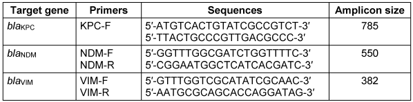
Statistical analysis
Categorical and continuous variables were expressed as frequencies. Multivariate regression analyses were used to estimate adjusted odds ratios (aORs) to assess the association between carbapenemase production and enterobacterales species, using MedCalc Version 23.0.2 (Ostend, Belgium). All analyses with p-values <0.05 at a 95% confidence interval (CI) were considered statistically significant.
Results
Identification of clinical enterobacterales isolates
The biochemical reactions of isolates, as determined by the API 20E System (Table 2 [Tab. 2]), showed that E. coli was the predominant (37%), followed by K. oxytoca 17%, K. pneumoniae 15% , Providencia spp. (10%) Citrobacter spp. (4%) and P. mirabilis (4%) (Table 2 [Tab. 2]). Based on sample type, urine samples had the highest recovery rate of enterobacterales (42%), followed by sputum (16%), wound swabs (12%), stool (12%) and blood cultures (5%) (Table 3 [Tab. 3]).
Table 2: Biochemical reactions profile specific for each enterobactarales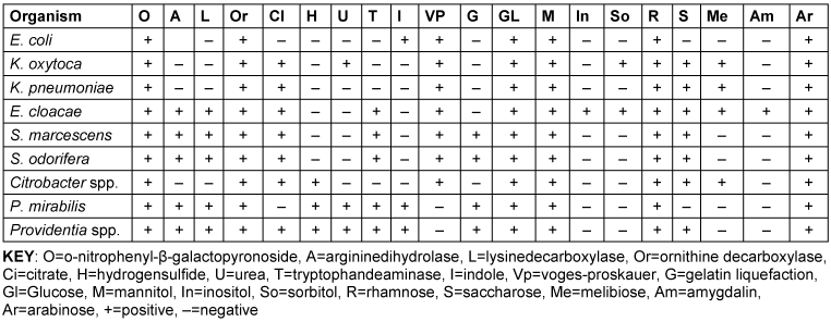
Table 3: Distribution and frequency of enterobacterales based on sample type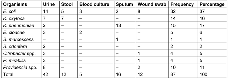
Frequency of CRE
The prevalence of carbapenem resistance among the enterobacterales isolates was 19.5% (17/87) (Table 3 [Tab. 3]). The distribution of isolates among species shows that E. coli accounts for the highest proportion (29%), followed by K. oxytoca (17%), K. pneumoniae, and S. odorifera (12%) (Table 3 [Tab. 3]). The remaining species, including Providencia spp., E. cloacae, Citrobacter spp., P. mirabilis and S. marcescens, each account for a smaller proportion (≤12%). Using the EDTA-modified Hodge test, the prevalence of CRE was 11.5%, and S. odorifera showed the highest carbapenem resistance (p=0.04; Table 4 [Tab. 4]).
Table 4: Prevalence of confirmed carbapenem-resistant strains by species of enterobacterales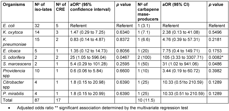
Antimicrobial susceptibility pattern of CRE
The pattern of resistance among carbapenemase producers to non-carbapenem agents in descending order of frequency: amoxicillin-clavulanate (70%), gentamicin (50%), ceftazidime (40%), ciprofloxacin (40%), and nitrofurantoin (20%) (Table 5 [Tab. 5]). Of the MDR carbapenemase-producers, blaNDM and blaVIM were detected in Serratia odorífera, E. cloacae, S. marcescens from urine samples and P. mirabilis and K. oxytoca from blood culture (Figure 1 [Fig. 1]).
Table 5: Antimicrobial resistance pattern of carbapenemase-producing enterobacterales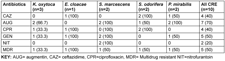
Figure 1: PCR Product of carbapenamase-encoding genes detected from enterobacterales on 1% (w/v) agarose gel.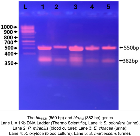
Discussion
Enterobacterales are a major component of the human gut; they are dynamic and play significant ecological roles in health and disease [23]. The prominent role of enterobacterales is their contribution to the spread of the AMR pandemic. The gut acts as a significant reservoir for these resistomes, such as carbapenemases, facilitating their transfer to other organs and tissues, leading to colonisation and subsequent infections (urinary tract infections, bloodstream infections) that are difficult to treat.
Reporting CRE from human samples is critically important for guiding patient treatment, preventing the spread of these “superbugs” within healthcare facilities and the community, and informing public health policy on AMR. It provides essential data to guide clinical decisions and prevent widespread transmission.
From the current study, E. coli (36.8%) was the predominant etiological agent of clinical infections. This corroborates numerous previous studies worldwide, such as the 35.1% reported in Nigeria (35.1%) [21], China (33.9%) [24], and Zambia (37.8%) [25]. However, higher prevalence values in Romania (41.5%) [26] and the Democratic Republic of Congo (58.9%) [27]. We reported a higher frequency of E. coli clinical isolates than those identified in Palestine (30.3%) and Ethiopia (14.2%) [28], [29]. These variances could be attributed to geographical disparities, technical differences, the procedures utilised, and the experience of the laboratory personnel who performed the investigations. Other important factors that could influence the recovery rate of E. coli include the specific sample type and patient population (e.g., healthy individuals vs. hospitalised patients). In this regard, urinary tract infections are the most common source, as corroborated by our findings.
Among enterobacterales species, carbapenem-resistant E. coli (CREc) was the most common (29.4%, 5/17), higher than the 7% global pooled prevalence of CREc [30]. Subregional comparison shows that CREc prevalence is higher than in high-income countries, such as Portugal, whereas CREc was as low as 0.5% among invasive isolates. It appears that CREc is endemic in Nigeria, as reflected by a recent study in Lagos, which reported a prevalence of 46% [31]. Moreover, the previous meta-analysis suggests that CRE is generally endemic in Nigeria [15]. Higher prevalence of CREc in human patients is primarily driven by healthcare-associated factors, including extensive antibiotic use, prolonged hospital stays, and the presence of invasive medical devices.
The 19.5% prevalence of CRE observed in this study was higher than the 14% reported in a global meta-analysis of CRE colonization [14]. The difference could be likened to the fact that enterobacterales from clinical infections have a greater capacity to acquire carbapenem resistance than in colonisation states due to antibiotic pressure [3]. It is important to note that some meropenem-resistant CRE were negative on the modified Hodge test, indicating they are non-carbapenemase producers. The key mechanisms responsible for carbapenem resistance in non-carbapenemase producers involve both chromosomal factors (such as point mutations in OprD protein) and non-chromosomal factors, including the production of non-carbapenemase β-lactamases [32], [33].
Worryingly, 40% of the MDR-CRE were simultaneously resistant to both carbapenems (CRE) and ceftazidime (a third-generation cephalosporin). This will ultimately result in limited treatment options, higher rates of treatment failure, increased patient mortality, longer hospital stays, and higher healthcare costs [34]. The blaNDM and blaVIM genes we detected in our CRE strains are among the major ones encoding enzymes that hydrolyze carbapenems. Remarkably, these CR genes are found in non-conventional (less common genera) enterobacterales, such as S. marcescens and Citrobacter spp., in clinical samples. Similar findings have been reported in recent studies globally [35], [36], [37]. These bacterial genera are predominantly environmentally adapted, highlighting the hospital environment as a vector and reservoir of carbapenem resistance. Recent studies globally have consistently highlighted the significant role of the hospital environment, particularly surfaces and plumbing systems (e.g., sink traps/drains), as key reservoirs and vectors for the transmission of carbapenem-resistant S. marcescens and Citrobacter spp. in clinical settings [38], [39].
Many antibiotic-resistant bacterial infections cause diseases, including pneumonia, which have been shown to contribute substantially to mortality [40], [41]. According to projections, there could be 1.91 million and 8.22 million AMR-related deaths worldwide in 2050 [41]. Therefore, the findings of the present study would help strengthen surveillance of CRE, which is critical to improving antimicrobial management and patient outcomes.
We explicitly acknowledge that the odds ratio analysis of small subgroups of isolates could limit the generalizability of the findings; hence, caution is warranted when interpreting the multivariate regression results. Whole-genome sequencing could be considered in future studies, as it provides detailed data on potential mobile genetic elements, transmission pathways, and relatedness among carbapenem-resistant strains.
Conclusion
Considering the global pooled prevalence of CRE of 14%, a high frequency was observed in the present study, with some also resistant to third-generation cephalosporins. Remarkably, the blaNDM- and blaVIM-genes were found in non-conventional enterobacterales, indicating their evolving roles as reservoirs of these critical priority AMR genes. A broader scope of surveillance, including environmental samples, is needed to understand the burden of CRE in healthcare settings. It is recommended that actionable plans prioritize enhanced genomic tracking, stringent infection control, rigorous diagnostic stewardship, and stricter antibiotic management to prevent further dissemination of these critical genes.
Notes
Authors’ ORCIDs
- Abubakar A: https://orcid.org/0009-0008-4171-4321
- Lawal G: https://orcid.org/0000-0001-7916-6000
- Abdullahi IN: https://orcid.org/0000-0002-5511-1272
Ethical approval
Ethical approval was obtained from the Ethics Review Committee of the Federal Teaching Hospital Gombe.
Funding
None.
Acknowledgments
Authors appreciate the technical support by the laboratory staff of Federal Teaching Hospital, Gombe, during the collection of enterobacteriales isolates.
Competing interests
The authors declare that they have no competing interests.
Availability of data and material
All data generated from this study are presented in the tables and figures; however, further requests could be made through the corresponding author.
AI usage statement
Artificial intelligence tool (PaperPal) was used only to improve the English language of the manuscript. All scientific contents, and references were reviewed, edited, and approved by the authors.
References
[1] Smith HZ, Hollingshead CM, Kendall B. Carbapenem-Resistant Enterobacterales. Treasure Island (FL): StatPearls Publishing; 2024 Feb 2. Available from: https://www.ncbi.nlm.nih.gov/books/NBK551704/[2] Wei Y, Luo H, Chen X, Yan Q. Epidemiology of subsequent carbapenem-resistant enterobacterales (CRE) infection among rectal carriers: A meta-analysis of incidence, risk factors and their association with geographic region and age. Int J Antimicrob Agents. 2024 Nov;64(5):107335. DOI: 10.1016/j.ijantimicag.2024.107335
[3] Alraddadi BM, Heaphy ELG, Alzahrani MS, Alqadi M, Qashqari MS, Alhuthali MS, Al Hroub MK, Hefni L, Alshukairi AN, Aldabbagh Y, Qutub M. Association Between Carbapenem-Resistant Enterobacterales (CRE) Colonization Status at Time of Hospital Admission and the Subsequent Development of CRE Infection and Mortality in High-Risk Patients. Infect Drug Resist. 2024 Oct 25;17:4655-64. DOI: 10.2147/IDR.S479487
[4] WHO. WHO Bacterial Priority Pathogens List, 2024: Bacterial pathogens of public health importance to guide research, development and strategies to prevent and control antimicrobial resistance. Geneva: World Health Organization; 2024.
[5] Ma J, Song X, Li M, Yu Z, Cheng W, Yu Z, Zhang W, Zhang Y, Shen A, Sun H, Li L. Global spread of carbapenem-resistant Enterobacteriaceae: Epidemiological features, resistance mechanisms, detection and therapy. Microbiol Res. 2023 Jan;266:127249. DOI: 10.1016/j.micres.2022.127249
[6] Gomez-Simmonds A, Annavajhala MK, Seeram D, Hokunson TW, Park H, Uhlemann AC. Genomic epidemiology of carbapenem-resistant Enterobacterales at a New York City hospital over a 10-year period reveals complex plasmid-clone dynamics and evidence for frequent horizontal transfer of blaKPC. Genome Res. 2024 Nov 20;34(11):1895-907. DOI: 10.1101/gr.279355.124
[7] European Centre for Disease Prevention and Control. Carbapenem-resistant Enterobacterales, third update – 3 February 2025. Stockholm: ECDC; 2025. Available from: https://www.ecdc.europa.eu/en/publications-data/carbapenem-resistant-enterobacterales-rapid-risk-assessment-third-update
[8] Yang X, Lou Z, Wang X, Li Z, Liu Q, Guo K, Yang Y, Gong L, Wang K, Xu H, Zheng B, Liu W, Fu C, Chen H, Jiang X. Resistance profile and influence factors of carbapenem-resistant Klebsiella pneumoniae (CRKP) causing infections in China: a systematic review and meta-analysis. Ann Clin Microbiol Antimicrob. 2025 Oct 14;24(1):56. DOI: 10.1186/s12941-025-00827-2
[9] Fasciana T, Antonelli A, Bianco G, Lombardo D, Codda G, Roscetto E, Perez M, Lipari D, Arrigo I, Galia E, Tricoli MR, Calvo M, Niccolai C, Morecchiato F, Errico G, Stefani S, Cavallo R, Marchese A, Catania MR, Ambretti S, Rossolini GM, Pantosti A, Palamara AT, Sabbatucci M, Serra N, Giammanco A. Multicenter study on the prevalence of colonization due to carbapenem-resistant Enterobacterales strains before and during the first year of COVID-19, Italy 2018-2020. Front Public Health. 2023 Dec 19;11:1270924. DOI: 10.3389/fpubh.2023.1270924
[10] Le HQN, Dang LML, Pham NTT, Van Le C, Lam TK, Luong LH, Huynh TH. Carbapenem resistance and carbapenemase classification of Enterobacteriaceae in intensive care unit: a cross-sectional study. Med Pharm Res. 2025 Aug 6;9(3):334–46. DOI: 10.32895/mpr.25.00001
[11] Elshamy AA, Aboshanab KM. A review on bacterial resistance to carbapenems: epidemiology, detection and treatment options. Future Sci OA. 2020 Jan 27;6(3):FSO438. DOI: 10.2144/fsoa-2019-0098
[12] Verma A, Singh N, Agarwal J, Sen M, Das A. Emergence and Spread of Carbapenem Antimicrobial Resistance: A Review on Mechanism of Drug Resistance and Laboratory Detection. Ann Afr Med. 2026 May 1;25(3):495-503. DOI: 10.4103/aam.aam_169_25
[13] Simner PJ, Pitout JDD, Dingle TC. Laboratory detection of carbapenemases among Gram-negative organisms. Clin Microbiol Rev. 2024 Dec 10;37(4):e0005422. DOI: 10.1128/cmr.00054-22
[14] Zhong Y, Huang J, Ning L, Xiong Y, Wu Y. The silent spread of resistance: Global patterns of CRE colonization across health care and community settings. Am J Infect Control. 2026 Jun;54(6):659-65. DOI: 10.1016/j.ajic.2025.12.003
[15] Irekeola AA, Shueb RH, Engku Abd Rahman ENS, Afolabi HA, Wada Y, Elmi AH, Hakami MA, Alghzwani SM, Elnoubi OA, Alshehri AA. High prevalence of carbapenem-resistant Enterobacterales (CRE) in human samples from Nigeria: A systematic review and meta-analysis. Heliyon. 2024 Jul 20;10(15):e34926. DOI: 10.1016/j.heliyon.2024.e34926
[16] Hassall J, Coxon C, Patel VC, Goldenberg SD, Sergaki C. Limitations of current techniques in clinical antimicrobial resistance diagnosis: examples and future prospects. NPJ Antimicrob Resist. 2024 Jun 17;2(1):16. DOI: 10.1038/s44259-024-00033-8
[17] Belkum van A. Next generation sequencing as a panacea for antibiotic susceptibility testing: yea or nay? Front Public Health. 2025 Aug 18;13:1650925. DOI: 10.3389/fpubh.2025.1650925
[18] Tristancho-Baró A, Franco-Fobe LE, Ariza MP, Milagro A, López-Calleja AI, Fortuño B, López C, Latorre-Millán M, Clusa L, Martínez R, Torres C, Rezusta A. Genomic Characterization of Carbapenemase-Producing Enterobacteriaceae from Clinical and Epidemiological Human Samples. Antibiotics (Basel). 2025 Jan 6;14(1):42. DOI: 10.3390/antibiotics14010042
[19] Lee YQ, Sri La Sri Ponnampalavanar S, Wong JH, Kong ZX, Ngoi ST, Karunakaran R, Lau MY, Abdul Jabar K, Teh CSJ. Investigation on the mechanisms of carbapenem resistance among the non-carbapenemase-producing carbapenem-resistant Klebsiella pneumoniae. Front Cell Infect Microbiol. 2024 Sep 18;14:1464816. DOI: 10.3389/fcimb.2024.1464816
[20] Yang Y, Li X, Sun L, Wang X-K, Zhang Y-W, Pang J, Li G-Q, Hu X-X, Nie T-Y, Yang X-Y, Liu J-H, Brandis G, You X-F, Li C-R. High level non-carbapenemase carbapenem resistance by overlaying mutations of mexR, oprD, and ftsI in Pseudomonas aeruginosa. Microbiol Spectr. 2025 Jan 7;13(1):e0139824. DOI: 10.1128/spectrum.01398-24
[21] Shitta G, Makanjuola O, Adefioye O, Olowe OA. Extended spectrum beta lactamase (ESBL), blaTEM,blaSHV and blaCTX-M, resistance genes in community and healthcare associated gram negative bacteria from Osun State, Nigeria. Infect Disord Drug Targets. 2021;21(4):595-602. DOI: 10.2174/1871526520999200729181559
[22] CLSI. Performance Standards for Antimicrobial Susceptibility Testing. 35th edn. CLSI Supplement M100. Clinical Laboratory Standard Institute; 2025.
[23] Moreira de Gouveia MI, Bernalier-Donadille A, Jubelin G. Enterobacteriaceae in the human gut: Dynamics and ecological roles in health and disease. Biology (Basel). 2024 Feb 23;13(3):142. DOI: 10.3390/biology13030142
[24] Long S, Zhong M, Huang X, Zhang J, Liu X, Yu H. Temporal shifts in etiological agents and trends in antimicrobial resistance of bloodstream infection in Southwest China from 2016 to 2023. Infect Drug Resist. 2025 Mar 11;18:1367-79. DOI: 10.2147/IDR.S514966
[25] Kasanga M, Shempela DM, Daka V, Mwikisa MJ, Sikalima J, Chanda D, Mudenda S. Antimicrobial resistance profiles of Escherichia coli isolated from clinical and environmental samples: findings and implications. JAC Antimicrob Resist. 2024 Apr 27;6(2):dlae061. DOI: 10.1093/jacamr/dlae061
[26] Goleanu Vasiloiu CD, Vrancianu CO, Goleanu DA, Tantu MM, Csutak O. Trends in antibiotic resistance of Escherichia coli strains isolated from clinical samples (2019-2023): A hospital-based retrospective analysis. Pathogens. 2025 Sep 13;14(9):927. DOI: 10.3390/pathogens14090927
[27] Irenge LM, Kabego L, Vandenberg O, Chirimwami RB, Gala JL. Antimicrobial resistance in urinary isolates from inpatients and outpatients at a tertiary care hospital in South-Kivu Province (Democratic Republic of Congo). BMC Res Notes. 2014 Jun 18;7:374. DOI: 10.1186/1756-0500-7-374
[28] Arman G, Zeyad M, Qindah B, Abu Taha A, Amer R, Abutaha S, Koni AA, Zyoud SH. Frequency of microbial isolates and pattern of antimicrobial resistance in patients with hematological malignancies: a cross-sectional study from Palestine. BMC Infect Dis. 2022 Feb 10;22(1):146. DOI: 10.1186/s12879-022-07114-x
[29] Kibret M, Abera B. Antimicrobial susceptibility patterns of E. coli from clinical sources in Northeast Ethiopia. Afr Health Sci. 2011;11:S40-5. DOI: 10.4314/ahs.v11i3.70069
[30] Walker MM, Roberts JA, Li Y, Sime FB. A systematic review and meta-analysis of the global epidemiology of carbapenem-resistant Escherichia coli. Int J Antimicrob Agents. 2025 Nov 17;67(1):107668. DOI: 10.1016/j.ijantimicag.2025.107668
[31] Salau M, Kositanont U, Noisumdaeng P, Ogunsola F, Ettu AO, Adewojo D, Ojimma C, Ojomaikre O, Changkaew K. Characterization of Carbapenem-Resistant Gram-Negative Bacilli Isolates in Multispecialty Private Hospitals in Lagos, Nigeria. Infect Dis Rep. 2025 Sep 21;17(5):119. DOI: 10.3390/idr17050119
[32] Manageiro V, Paiva JA, Caniça M. Vigilância da resistência aos antibióticos em Portugal de 2015 a 2022. Bol Epidemiol Obs. 2023;12:39-45. Available from: http://hdl.handle.net/10400.18/8747
[33] Miftode IL, Radu VD, Jigoranu RA, Leca DA, Prepeliuc CS, Pasare MA, Miftode RS, Grigoriu MG, Parângă TG, Miftode EG. Novel Insights into Carbapenem Resistance: Mechanisms, Diagnostics, and Future Directions. Antibiotics (Basel). 2026 Mar 5;15(3):270. DOI: 10.3390/antibiotics15030270
[34] Alshehail BM, Alwezzeh MJ, Almalki HH, Alnimr A, Wali H, Al Jamea Z, Al Rashed AS, Alhajri M, Abdulaal HA, Alanbari LA, Alsowaida YS, Alamri A, Almuthen S, Azaiez F, Alzahrani S, Zakari N, Asiri J, Alanazi W, Bakkar M, Alfifi A, Alzuwayed O, El-Saed A, AlBahrani S. Recurrence, Readmission, and Key Mortality Predictors in Patients with Carbapenem-Resistant Enterobacterales Infections. Diagnostics (Basel). 2025 Nov 21;15(23):2957. DOI: 10.3390/diagnostics15232957
[35] Shugart A, Mahon G, Huang JY, Karlsson M, Valley A, Lasure M, Gross A, Pattee B, Vaeth E, Brooks R, Maruca T, Dominguez CE, Torpey D, Francis D, Bhattarai R, Kainer MA, Chan A, Dubendris H, Greene SR, Blosser SJ, Shannon DJ, Jones K, Brennan B, Hun S, D'Angeli M, Murphy CN, Tierney M, Reese N, Bhatnagar A, Kallen A, Brown AC, Spalding Walters M. Carbapenemase production among less-common Enterobacterales genera: 10 US sites, 2018. JAC Antimicrob Resist. 2021 Sep 4;3(3):dlab137. DOI: 10.1093/jacamr/dlab137
[36] Sabtcheva S, Stoikov I, Ivanov IN, Donchev D, Lesseva M, Georgieva S, Teneva D, Dobreva E, Christova I. Genomic Characterization of Carbapenemase-Producing Enterobacter hormaechei, Serratia marcescens, Citrobacter freundii, Providencia stuartii, and Morganella morganii Clinical Isolates from Bulgaria. Antibiotics (Basel). 2024 May 16;13(5):455. DOI: 10.3390/antibiotics13050455
[37] Mauffrey F, Bertelli C, Greub G, Grandbastien B, Senn L, Blanc DS. Plasmid diversity of Serratia marcescens and Klebsiella pneumoniae isolates involved in two carbapenem-resistant enterobacteriaceae outbreaks in a Swiss hospital. Microbiol Spectr. 2025 Jul;13(7):e0328424. DOI: 10.1128/spectrum.03284-24
[38] Cirkovic I, Brkic S. Beyond the spotlight: Enterobacter spp. as overlooked carbapenemase producers in Europe. Antibiotics (Basel). 2025 Oct 18;14(10):1045. DOI: 10.3390/antibiotics14101045
[39] McCallum GE, Hall JPJ. The hospital sink drain microbiome as a melting pot for AMR transmission to nosocomial pathogens. NPJ Antimicrob Resist. 2025 Jul 29;3(1):68. DOI: 10.1038/s44259-025-00137-9
[40] Kanani J. Autopsy analysis of sudden deaths in adults: Causes and demographics from a one-year prospective study. Curr Health Sci J. 2025 Jul-Sep;51(3):343-9. DOI: 10.12865/CHSJ.51.03.05
[41] GBD 2021 Antimicrobial Resistance Collaborators. Global burden of bacterial antimicrobial resistance 1990-2021: a systematic analysis with forecasts to 2050. Lancet. 2024 Sep 28;404(10459):1199-226. DOI: 10.1016/S0140-6736(24)01867-1
[42] Mohammed Y, Zailani SB, Onipede AO. Characterization of KPC, NDM and VIM type carbapenem resistance in enterobacteriaceae from North Eastern, Nigeria. J Biosci Med. 2015 Jan 1;03(11):100-7. DOI: 10.4236/jbm.2015.311013