Ulnar polydactyly
Sterre ten Berge 1
1 Department of Plastic, Reconstructive and Hand Surgery, Erasmus MC – University Medical Centre Rotterdam, Rotterdam, Netherlands
Abstract
Ulnar polydactyly is among the most common congenital hand anomalies. According to Temtamy and McKusick, it is classified as type A, a well-formed digit with tendons, bones and neurovascular structures, or type B, a rudimentary pedunculated underdeveloped digit. The condition results from genetic mutations affecting the radial-ulnar axis, often due to GLI3-mutations. It can present as an isolated anomaly or as part of various syndromes. Epidemiological groups show different clinical aspects and type B is the prevalent form overall. The diagnosis is primarily clinical, however, it can be supported by radiological imaging and referral to a clinical geneticist if needed. The goal of therapy is removal of the extra digit while preserving the function and sensation to the remaining digit. Type A-digits are surgically treated via various approaches, depending on their articulation and anatomy, and current evidence reveals good outcomes. Type-B digits can be managed via ligation, via sutures or clips, or via surgical excision, under local or general anesthesia. Surgical excision shows the lowest complication rate, although high-level comparative evidence is still limited.
Introduction
Polydactyly represents one of the most common congenital anomalies of the hand [1]. Supernumerary digits in ulnar polydactyly are classified as type A or type B, as described by Temtamy and McKusick [2]. These digits are typically non-functional and can interfere with daily life activities. The goal of surgical treatment is to improve hand function and aesthetic appearance.
Basics
Anatomy
The extra digits follow the classification system by Temtamy and McKusick into type A and type B forms [2].
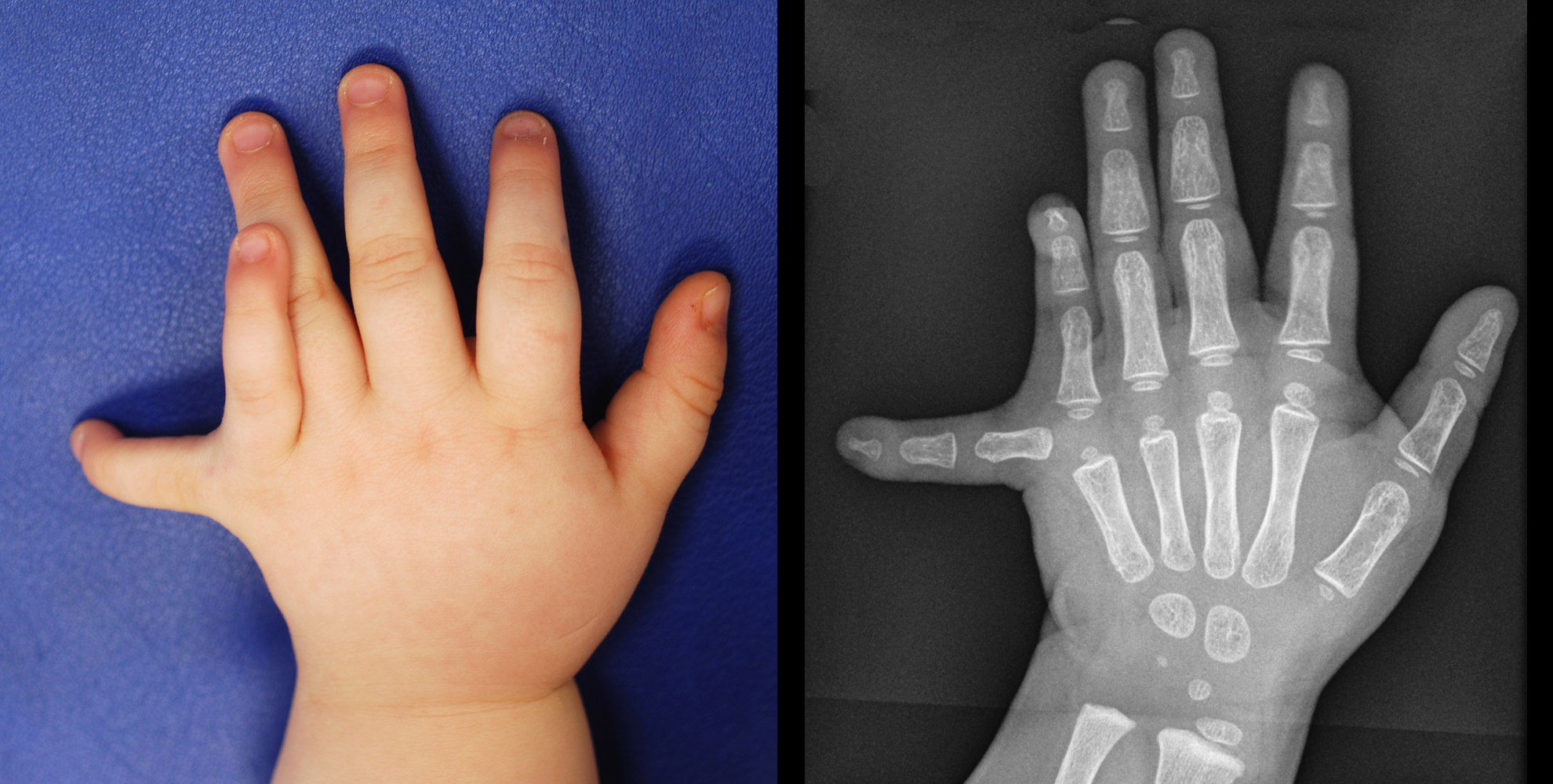
Type A ulnar polydactyly presents as a well-developed digit with variable anatomy, located on the ulnar border of the little finger. These digits articulate with either the fifth or a duplicated metacarpal, and the angle of articulation can range from less than 30° to 180° (Figure 1). Flexor and extensor tendons, and neurovascular structures are typically present [2]. These tendons can be anomalous and include the abductor digiti minimi (ADM) and the flexor digiti minimi brevis (FDMB). The interphalangeal joints are usually stiff and hypoplastic.
In case of duplication at the level of the MCP, whether due to a duplicated proximal phalanx articulating with a common MCP joint or bifid metacarpal, the ulnar collateral ligament attaches to the additional little finger. A duplicated metacarpal can contain the insertions of the opponens digiti minimi (ODM) and can be surrounded by the muscle bellies of the ADM and FDMB muscles.
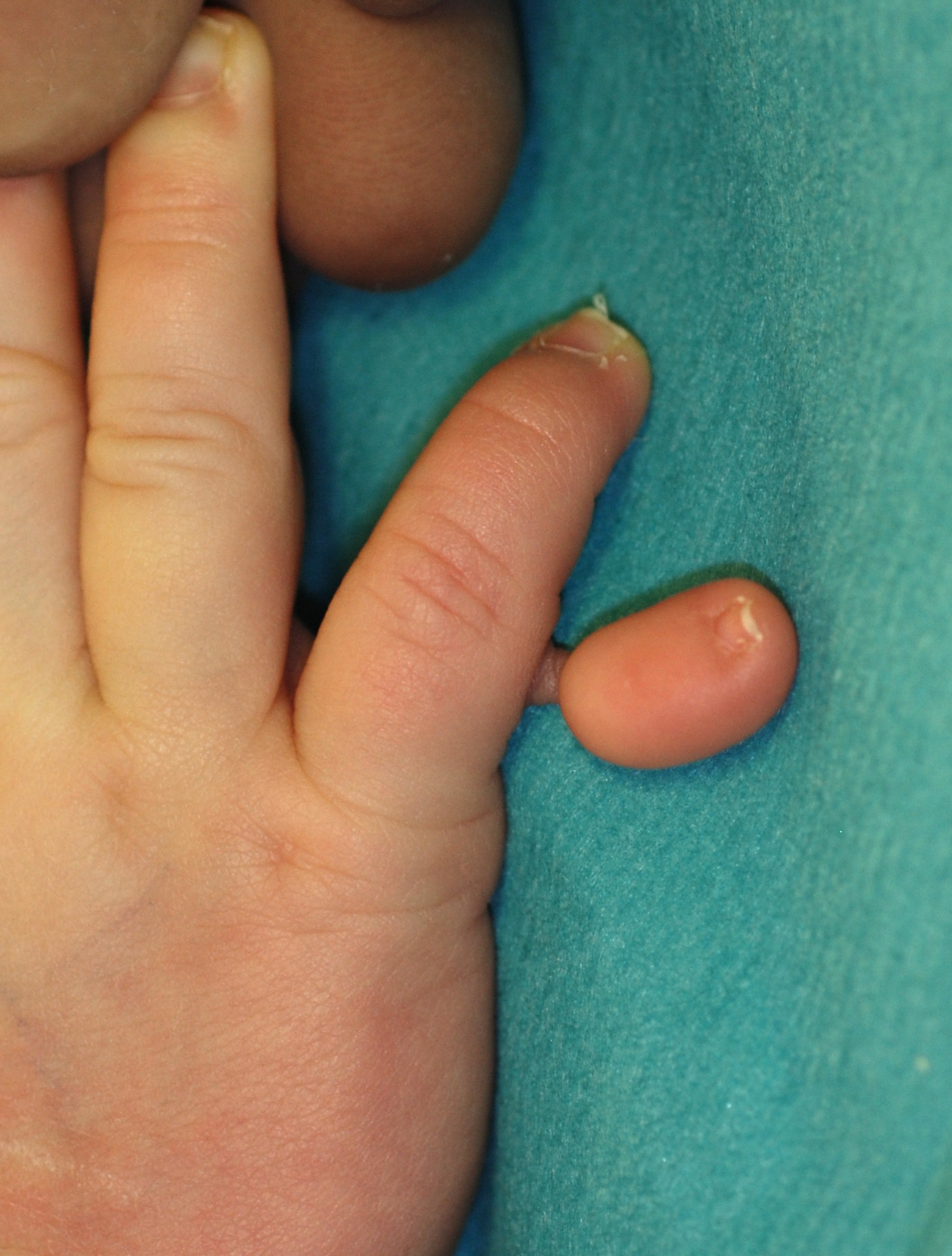
Type B ulnar polydactyly demonstrates a digital remnant, attached on the ulnar side of the fifth proximal phalanx (Figure 2). The anatomical structure of the digits varies from a small wartlike lump to a partially developed digit containing fibrocartilaginous ossicle and a hypoplastic nail, typically pedunculated and connected via a soft skin bridge [2]. These digits are supplied via a small neurovascular structure.
Embryology
Ulnar polydactyly results from a developmental error in the radial-ulnar axis. Genetic mutations, e.g. GLI3, affecting these pathways can cause ulnar polydactyly [1], [3], [4]. GLI3 proteins exist in active forms (GLI3A) and in repressor forms (GLI3R). The normal balance of GLI3 is disturbed when GLI3R increases relatively compared to GLI3A. It is suggested that this disturbance of the normal balance possibly contributes to the formation of ulnar polydactyly [5].
Epidemiology
Ulnar polydactyly is among the most common congenital hand anomalies. Type B ulnar polydactyly is the prevalent form overall, occurring approximately 1 in 150 live births in African Americans and up to 1 in 1,300 in the Caucasian population [6]. Syndromal disorders are more commonly seen with Type A [7].
Clinical aspects differ between ethnic groups. In patients of African descent, it presents most commonly as type B and is often bilateral (70%). The condition affects males and females equally, with the left hand more commonly affected [8]. Ulnar polydactyly is usually inherited in an autosomal dominant way and occurs mostly isolated. It is rarely associated with other hand anomalies, congenital syndromes, or non-syndromal systemic abnormalities.
Clinical aspects differ between ethnic groups. In patients of African descent, it presents most commonly as type B and is often bilateral (70%). The condition affects males and females equally, with the left hand more commonly affected [8]. Ulnar polydactyly is usually inherited in an autosomal dominant way and occurs mostly isolated. It is rarely associated with other hand anomalies, congenital syndromes, or non-syndromal systemic abnormalities.
In patients of non-African descent, ulnar polydactyly tends to occur sporadically. Only around 5% of non-Africans demonstrate a recognizable inheritance pattern. Types A and B occur with similar frequency, but bilateral involvement is less common (20%), and the condition is more prevalent in males. Associated hand conditions are more likely, and foot involvement, such as fibular toe polydactyly, can also be present. Additionally, it may be associated with other congenital syndromes with either autosomal dominant or autosomal recessive patterns of inheritance (see Table 1) [9].
|
Origin |
Inheritance |
Clinical aspects |
|
|
Greig syndrome |
GLI3 gene (7p14.1) |
Autosomal dominant |
Frontal bossing with macrocephaly, hypertelorism, radial polydactyly of at least one limb, radial- and ulnar polydactyly of the other limbs and cutaneous syndactyly. |
|
Ellis–van Creveld syndrome |
EVC and EVC2 gene (4p16)
|
Autosomal recessive |
Strabismus, partial cleft lip, disproportionate short stature (long, narrow thorax with shortening of all extremities), ulnar polydactyly (usually bilateral, sometimes unilateral or feet involvement), hidrotic ectodermal dysplasia (hypoplastic nails, fine hair and partial anodontia, enamel hypoplasia, multiple frenula), epi- and hypospadias, cryptorchidism. |
|
Bardet-Biedl syndrome |
BBS1 gene (11q13.2) and BBS10 gene (12q21.2)
|
Autosomal recessive |
Strabismus, astigmatism, cataract, oral and/or dental abnormalities (e.g. crowding teeth, hypodontia, high-vaulted palate), ulnar polydactyly, cognitive impairment, obesity and hypogonadism. |
|
Joubert syndrome |
Various genes involved. Two loci on chromosomes 9q34 (INPP5E) and 11p12-q13 (TMEM216)) |
Autosomal recessive |
Abnormal eye movements (e.g. oculomotor apraxia (OMA) or nystagmus), head thrusting or horizontal head nodding (e.g. a ‘no- no” head tremor) to compensate for OMA, ulnar polydactyly, hypotonia and abnormal respiratory pattern (alternating apnea and/or tachypnea). |
|
Ulnar mammary syndrome |
TBX3 gene (12q24.21) |
Autosomal dominant |
Broad facial structure ending in a defined chin, broad nasal tip, wide nasal base, bifid tongue tip, hypodontia, hypoplastic or absent humerus, hypoplastic or absent ulna and/or radius, hypoplastic distal phalanges of the fifth finger(s), absent ulnar ray digits (3rd, 4th, 5th rays), camptodactyly, ulnar polydactyly, cryptorchidism, areolar or nipple hypoplasia, obesity, and short stature. |
|
Carpenter syndrome |
RAB23 gene (6p12.1) and MEGF8 gene (19q13.2) |
Autosomal recessive |
Craniosynostosis, epicanthal folds, down-slanting palpebral fissures, flat nasal bridge, low-set, posteriorly rotated malformed ears, pre-auricular tags, undeveloped maxilla and mandible, ulnar polydactyly (hands) with broad thumbs or absent middle phalanges, clinodactyly, camptodactyly, tibial polydactyly (feet), cutaneous syndactyly, brachydactyly, obesity, macrosomia, umbilical hernia, hypogonadism and cryptorchidism. |
|
Acrocallosal syndrome |
KIF7 (15q26.1) and, rarely, GLI3 (7p14.1) |
Autosomal recessive |
Macrocephaly with prominent forehead and occiput, hypertelorism, large anterior fontanel, broad nasal bridge, and short mandible, triangular mouth, cleft lip, cleft palate, higharched palate, brachydactyly, radial polydactyly, ulnar polydactyly, syndactyly of the feet, imperforate anus, hypospadia, cryptorchidism and umbilical hernia. |
Classification
The OMT classification describes ulnar polydactyly (IB2vi) as a malformation in the hand plate following the radio-ulnar axis [10]. Within this condition, supernumerary digits are further categorized as type A or type B, as described by Temtamy and McKusick [2]. Type A describes a fully developed digit, articulating with either the fifth metacarpal or a duplicated metacarpal. Type B describes an insufficiently developed digit, formed either as a small bump or as a hypo-plastic digit pedicled on a small stalk.
Clinical aspects
Type A polydactyly requires examination of the flexion and extension tendons. Lack of creases over the interphalangeal joints indicates the disfunction of these tendons [11]. Type B polydactyly is attached solely via soft tissue. The width of the pedicle is decisive in determining whether clipping is possible or resection is required.
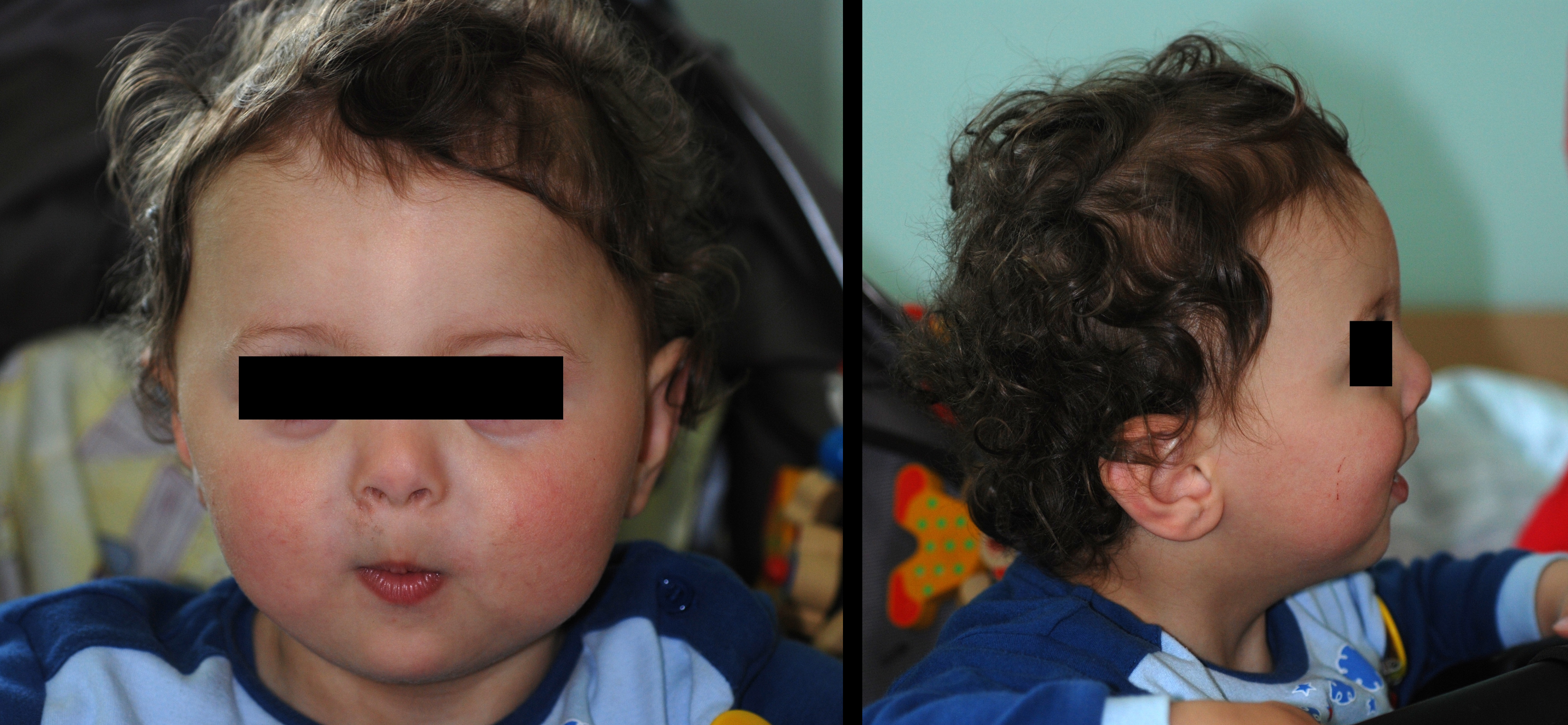
Given the potential syndromic associations, physical examination should also focus on syndromal features (see Table 1). Examination of the lower limbs should be included as fibular toe polydactyly might be present [5]. Greig syndrome is the most common, caused by mutations in the GLI3-gene and inherited in an autosomal dominant way. Features of Greig syndrome include frontal bossing with macrocephaly, hypertelorism, radial polydactyly of at least one limb, radial- and ulnar polydactyly of the other limbs and cutaneous syndactyly (Figure 3).
Diagnostics
Ulnar polydactyly is primarily a clinical diagnosis. Type B supernumerary digits generally do not necessitate further diagnostic investigation. For type A cases, radiological imaging helps assessing the articulation between the supernumerary digit and the fifth digit, which is important for the surgical approach [12] (Figure 4). Given the potential for inheritance, family history should be obtained.
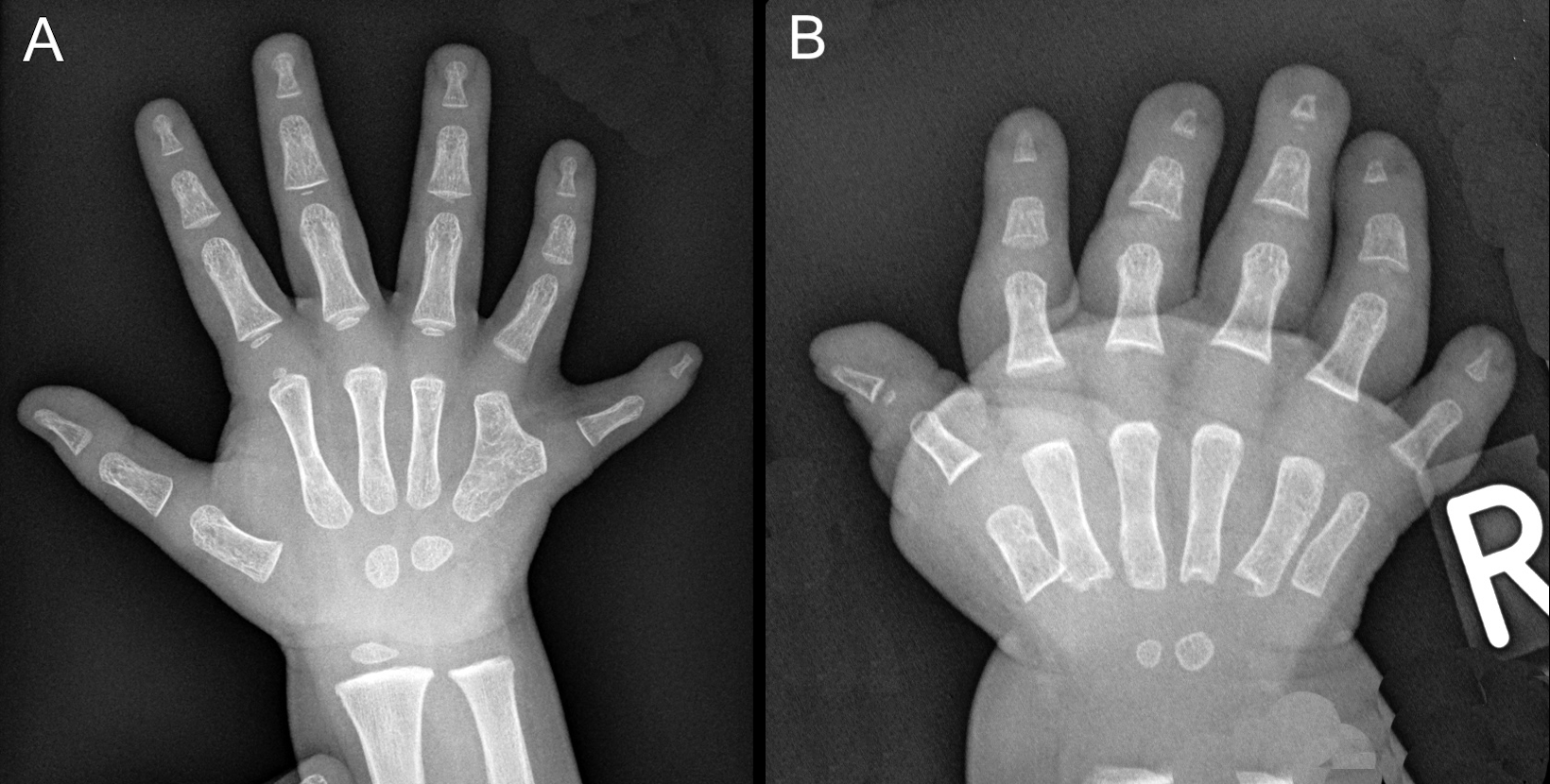
If clinical findings suggest syndromic involvement, referral to a clinical geneticist is recommended. Early genetic testing can help establish a diagnosis, which provides insights into giving adequate treatment. It also plays a critical role in identifying other family members who may be at risk and for supporting future family planning.
Therapy
The aim of surgical treatment is to remove the extra digit while preserving the function and sensation to the remaining digit.
For type A cases containing a common metacarpal articulation, an incision in the glabrous line is made at the base of the extra digit, which can be extended proximal and distal to expose the ADM. Then, the ulnar collateral ligament and ADM insertions are elevated, and the duplicated flexor and extensor tendons (often hypoplastic) can be assessed to the accessory digit. If the tendons to the little finger/fifth ray are sufficient, the extra tendons can be transected. The neurovascular bundle to the extra digit is cut off. If the articular surface is too wide, the head of the metacarpal bone is narrowed by longitudinal osteotomy. The collateral ligament and ADM are reinserted on the remaining proximal phalanx’ base.
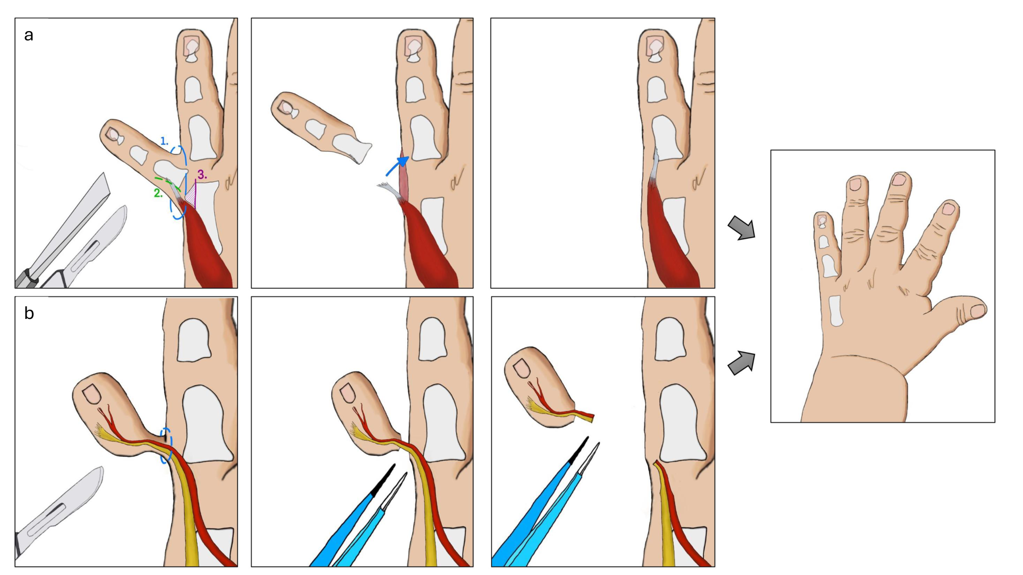
In the presence of a duplicated metacarpal, the supernumerary digit is amputated in a standard ray fashion, with transfer of the ADM to the preserved little finger (Figure 5a). A racket incision is used around the extra ray, leaving a scar along the glabrous line. In bilateral cases, both hands are operated on simultaneously to minimise anaesthetic use. If for logistical reasons both hands have to be operated on one after another, then approaching the easier side first may help in anticipating surgical requirements and planning for the more challenging extra digit [13]. However, starting with the dominant side has also been advocated, so the patient has the dominant side available at the second operation at an older age.
For type A polydactyly, long term treatment outcomes seem favorable as most patients were pleased with postoperative results, according to Cordray et al. [14]. Satisfaction scores for hand appearance were high, although patients thought their hands could look better. Hand function, following the PROMIS Upper Extremity Score, was below pediatric standards, particularly for older children. The complexity of polydactyly seems to correlate with long-term sequelae.
For type B cases, there are different opinions regarding the optimal age and method of treatment. Options for treatment include ligation, via suture or vascular clips, or resection, via local or general anesthesia (Figure 5b).
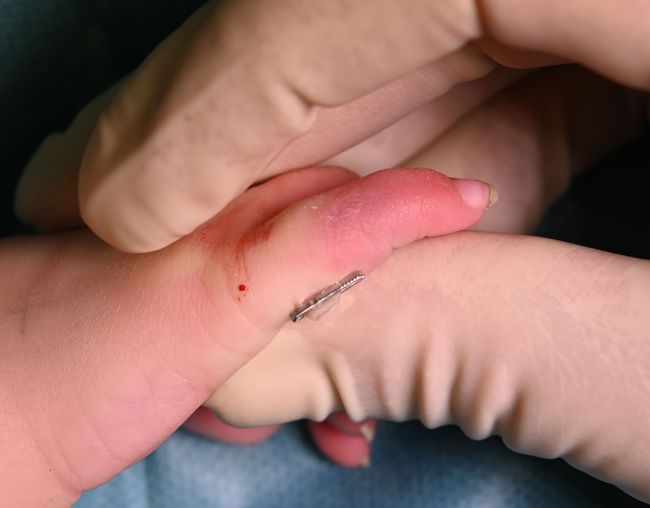
Suture ligation is an old method where midwives used to tie off the floating digit without sedation, allowing it to become necrotic and fall off. This approach is still being used today. Vascular clip ligation and resection can be performed when the clip is wide enough to fit around the base of the extra digit, thereby minimizing the risk of bleeding. This treatment is inexpensive and straightforward to fulfil. However, suture ligation can lead to unsightly nubbins and painful neuromas [15]. Clipping at the very base of the supernumerary digit can lead to smaller, flatter nubbins than with ligating with a suture, which might result in lower revision rates [16] (Figure 6). Regarding timing of treatment, ligation and clipping can be performed at a young age shortly after birth [17].
Surgical excision could result in higher patient satisfaction and lower revision rates for persistent nubbins and painful neuroma [18]. Surgery can be conducted under local or general anesthesia. Regarding excision under local anesthesia, removal of the supernumerary digit can be executed with minimal distress in an operating room without need for general anesthesia under “milk sedation”, i.e. the baby is fed just before or at the beginning of the procedure. This procedure is most suitable for babies less than 2–3 months old. Beyond 3 months of age, they are lively and it will be difficult to operate under local anesthetic. Therefore, formal surgical excision is usually delayed until the age of 12 months regarding safer general anesthesia [19].
The options for treatment can vary amongst surgeons and there is little literature comparing outcomes. To illustrate, a systematic review examining 900 articles could only include 10 and was not conclusive [20]. The authors mention a complication rate of 23.5% for ligation compared with 3% for excision. Efforts by the authors to conduct a multicenter randomized controlled trial (RCT) were unsuccessful due to a lack of consensus among surgeons regarding the optimal approach.
The currently preferred method
Excision under local anesthesia is mostly preferred amongst pediatric hand surgeons in most of Western Europe. For this procedure, experienced support staff, diathermy, good light, and mobile seating are needed. The baby needs to be fed with bottle milk or sucrose water before or while it takes place.
When preparing the skin, cover the baby’s eyes with a swab, especially when alcohol based. The baby should be swaddled by the attending nurse, positioning the hand. Once the baby is asleep, prewarmed local anesthetic with adrenaline can be administered. Inject the anesthetic proximal to the extra digit. Start with volar incision and follow with dorsal incision just through the dermis ensuring sufficient tissue present for good contour. Do not apply traction to the extra digit. Use bipolar diathermy to the vessel and nerve. Close with interrupted absorbable sutures, excising dog ears and covering with narrow, self-adhesive plaster strips for closing small wounds and self-adhesive gauze for securing wound dressings
Author's preferred method
Vascular clip ligation is performed until the age of three months (Figure 6). Anesthesia related risks are avoided, it takes relatively little time, and follow-up in the outpatient clinic is normally not necessary.
First, the nurse applies EMLA cream to the extra digit and its base, covers it with foil and a cotton glove, and after 30–45 minutes the bandages are removed. The EMLA dosage must be strictly followed since lidocaine- or prilocaine-containing substances are associated with a risk of methemoglobinemia, especially in young children. One parent holds the baby while the nurse positions the hand. The surgeon places two clips, based on the length of the pedicle, at its base and stacks it to prevent slipping, then removes the extra digit. Adhesive tapes and bandages are applied to secure the clips. Pain is typically minimal and occurs mainly during the placement of the clips and digit removal, subsiding shortly afterwards. The parents are contacted by phone for a follow-up a few weeks after the procedure, and an outpatient visit is scheduled if any complications arise.
Conclusion
As one of the most common congenital anomalies of the hand, ulnar polydactyly presents considerable variation in clinical presentation, epidemiology, and treatment strategies. Although most cases are of non-syndromic origin, a syndromic cause must always be considered, particularly in the presence of additional anomalies or in non-African populations. Diagnosis is clinical, though radiological imaging or referral to a clinical geneticist may be indicated in some cases. Surgical treatment depends on the anatomical structures in type A and of the pedicle width in type B. Patient age and parental preferences should be considered when deciding for treatment. High level evidence comparing techniques is still limited.
References
[1] Kyriazis Z, Kollia P, Grivea I, Stefanou N, Sotiriou S, Dailiana ZH. Polydactyly: Clinical and molecular manifestations. World J Orthop. 2023 Jan 18;14(1):13-22. DOI:10.5312/wjo.v14.i1.13[2] Temtamy S, McKusick VA. The genetics of hand malformations. Birth Defects Orig Artic Ser. 1978;14:364-92
[3] Dy CJ, Swarup I, Daluiski A. Embryology, diagnosis, and evaluation of congenital hand anomalies. Curr Rev Musculoskelet Med. 2014 Mar;7(1):60-7. DOI:10.1007/s12178-014-9201-7
[4] Seiler, JG; American Society For Surgery Of The Hand. Essentials of hand surgery. Philadelphia: Lippincott Williams & Wilkins; 2002
[5] Holmes LB, Nasri H, Hunt AT, Toufaily MH, Westgate MN. Polydactyly, postaxial, type B. Birth Defects Res. 2018 Jan;110(2):134-41. DOI:10.1002/bdr2.1184
[6] Weber, D. Baby-friendly and safe excision of postaxial polydactyly type B. IFFSH Ezine. 2018 May;8(2):26-9. Available from: https://www.ifssh.info/
[7] Bubshait DK. A review of polydactyly and its inheritance: Connecting the dots. Medicine (Baltimore). 2022 Dec 16;101(50):e32060. DOI:10.1097/MD.0000000000032060
[8] Hiro ME, Gottschalk HP, Light TR. Ulnar Polydactyly and Ulnar Dimelia. In: Laub DR, editor. Congenital Anomalies of the Upper Extremity. Cham: Springer Nature; 2021. p 351-366. DOI:10.1007/978-3-030-64159-7_21
[9] Ezaki M, James M, Wall L, Bauer A. The Care of Congenital Hand Anomalies. 3rd Edition. American Society for Surgery of the Hand; 2022
[10] Goldfarb CA, Ezaki M, Wall LB, Lam WL, Oberg KC. The Oberg-Manske-Tonkin (OMT) Classification of Congenital Upper Extremities: Update for 2020. J Hand Surg Am. 2020 Jun;45(6):542-7. DOI:10.1016/j.jhsa.2020.01.002
[11] Wolfe SW, Hotchkiss RN, Pederson WC, Kozin SH, Cohen MS, Al E. Green's operative hand surgery. Philadelphia: Elsevier, Cop; 2017
[12] Jones J, Weerakkody Y, Niknejad M, Yap J. Polydactyly. Radiopaediaorg. 2010 Sep 27 [cited 2024 Dec 1]. DOI:10.53347/rID-10844
[13] Watt AJ, Chung KC. Duplication. Hand Clin. 2009;May;25(2):215–27. DOI:10.1016/j.hcl.2009.01.001
[14] Cordray H, Shah AS, Buttrick E, Chang B, Little KJ, Al-Muhtaseb T, et al. Postoperative and long-term patient-reported outcomes of type A ulnar polydactyly. Plast Reconstr Surg. 2025;156(4):521e-33e. DOI:10.1097/PRS.0000000000012052
[15] Ganju N, Mantilla-Rivas E, Martinez PF, Manrique M, Escandón JM, Shah S, et al. A Retrospective review of patient-reported outcomes after postaxial polydactyly ligation and surgical excision. Plast Reconstr Surg Glob Open. 2024;12(1):e5557–7. DOI:10.1097/GOX.0000000000005557
[16] Mills JK, Ezaki M, Oishi SN. Ulnar polydactyly: long-term outcomes and cost-effectiveness of surgical clip application in the newborn. Clin Pediatr (Phila). 2014 May;53(5):470-3. DOI:10.1177/0009922813514479
[17] Zhou G, Dai L, Zhu J, Miao L, Wang Y, Liang J, et al. Epidemiological analysis of polydactylies in Chinese perinatals. Sichuan da xue xue bao Yi xue ban = Journal of Sichuan University Medical science edition. 2004 Sep;35(5):708-10
[18] Goebel GJ, Dawson S, Loewenstein SN, Adkinson JM. Long-Term Outcomes After Treatment for Type B Ulnar Polydactyly. J Pediatr Orthop. 2022 May-Jun 01;42(5):e466-e9. DOI:10.1097/BPO.0000000000002106
[19] Davidson A, Vutskits L. The new FDA drug safety communication on the use of general anesthetics in young children: what should we make of it? Pediatric Anesthesia. 2017 Mar 16;27(4):336-7. DOI:10.1111/pan.13122
[20] Chopan M, Sayadi L, Chim H, Buchanan PJ. To tie or not to tie: A systematic review of postaxial polydactyly and outcomes of suture ligation versus surgical excision. Hand (NY). 2020 May 1;15(3):303–10. DOI:10.1177/1558944718810885